Download
1 / 56
600 likes | 739 Vues
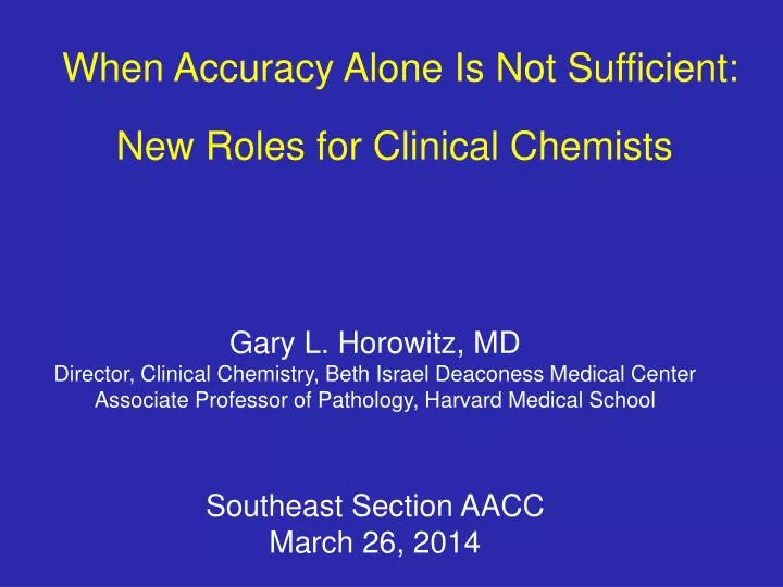
When Accuracy Alone Is Not Sufficient: New Roles for Clinical Chemists. Gary L. Horowitz, MD Director, Clinical Chemistry, Beth Israel Deaconess Medical Center Associate Professor of Pathology, Harvard Medical School. Southeast Section AACC March 26, 2014. Roles for Clinical Chemists.

E N D
When Accuracy Alone Is Not Sufficient:New Roles for Clinical Chemists Gary L. Horowitz, MD Director, Clinical Chemistry, Beth Israel Deaconess Medical Center Associate Professor of Pathology, Harvard Medical School Southeast Section AACC March 26, 2014
Roles for Clinical Chemists • Traditional Roles • Choose (and validate) instruments/methods • Generate accurate, timely results • Follow doctors’ “orders” • Plus potentially others
Roles for Clinical Chemists • New Roles: • Help implement clinical guidelines • HCV Ab testing for all Baby Boomers • Will detect 75% of undiagnosed cases (3M in US) • HCV (unlike HBV and HIV) is a curable disease • Explain potentially confusing tests • opiate immunoassay does not detect oxycodone(or methadone) • with every result – not just if you’re called • Help explain costs and TAT • HBV viral load unnecessary for diagnosis of HBV • it’s roughly 20 to 50-fold more expensive than HBsAg • Traditional Roles • Choose (and validate) instruments/methods • Generate accurate, timely results • Follow doctors’ “orders” • Plus potentially others 3
Numerous Other Examples • Immunoassays for testosterone • adequate for screening adult males for hypogonadism • Vitamin D (25 OH Vitamin D) • does your assay detect D2 at 100%? • eliminating orders for 1,25 Dihydroxy Vitamin D • Ethylene glycol or methanol: OK to test, but treat presumptively • “4th Generation HIV Ab”: Is it really helpful? • Immunosuppressants (CsA, Tacro, Rapa): • do your physicians know results are method-dependent? • it may be a “Send-Out”, but you can (should?) help • ESR: why are most of our labs still offering it? • if may not be done in your lab, but you can (should?) help • Glucose meter accuracy: • does it allow for tight (or even semi-tight) glycemic control? • do critical values need to be confirmed by central lab? • it may be not under your jurisdiction, but you can (should?) help
Three Examples TodayAll Illustrate Why Clinical ChemistsAre In a Strategic Position to Improve Care • Urine Dipstick Protein • a genuinely awful test: we need to eliminate it • or, at a minimum, highlight its deficiencies • Urine Albumin • Extremely underutilized test • Screening for CKD, an epidemic • Hemoglobin A1c • more complicated than you’d think • a poor surrogate for fingerstick glucose • time permitting, we may mention fructosamine and glycated albumin 5
Dipstick Urine Protein • among most common lab tests done • lab tests: diagnostic vs screening • discourage its use for screening • not simply wasteful (actually, it’s very inexpensive) • rather, potentially misleading
Case Scenario Dr. Jones screens all his diabetic patients by sending urine samples to your lab for dipstick proteins. As long as the dipstick is reported as negative, he is reassured that he has ruled out early diabetic nephropathy. Sounds reasonable, doesn’t it?
Proteinuria Physiology • virtually all proteins are too large to be filtered through a healthy glomerulus • once proteins do leak, there is no mechanism to reabsorb them • urine protein concentration reflects amount leakedpluswater content of urine , which varies with hydration • provides rationale for reporting urine protein not simply as concentration but as 24o collection
Protein/Creatinine Ratio • creatinine filtered through glomerulus • largely unsecreted and unreabsorbed by tubules • thus, its urine concentration reflects amount filteredpluswater content of urine, which varies with hydration • if you divide [protein]urine by [creatinine]urine, since water content of urine is in denominator of both, you eliminate the effect of hydration status • urine protein/creatinine ratio is an excellent surrogate for 24o urinary protein and can be done on any spot/random urine!
[protein]u is misleading • what can happen when you rely on [protein] alone • NB: conventional chemistry assay is no better !!
[protein]u is misleading • what can happen when you rely on [protein] alone • NB: conventional chemistry assay is no better !!
False Negative Type 1 • Bence-Jones Protein (BJP) • monoclonal free light chains • by definition, very small (23 kD) • so small, filtered by normal glomerulus (even without albuminuria!!) • not detected by dipstick method Adapted from Burtis, CA & Ashwood, ER. Tietz Fundamentals of Clinical Chemistry (4th Edition). Philadelphia: W.B Saunders, 1996, p.135.
Urine Protein Methods • dipstick: • method: protein error of pH indicators (c1909) • detects albumin > globulin > BJP • conventional chemistry assay: • method: denature protein, then detect resulting turbidity using spectrophometry • sensitive to all proteins, including BJP If a sample is dipstick negative, chemistry positive, it’s probably BJP • (micro)albumin: • method: immunoassay • detects only albumin
False Negative Type 2 • dipstick protein is not sensitive enough to rule out pathologic levels of proteinuriacannot distinguish low levels from 0 • definition: • analytic sensitivity = how low you can go? • assays for which sensitivity is particularly important: • TSH (3rd generation) • CRP (“hs-CRP”) • Troponin • D-Dimer • and, yes, urine protein!
Is Sensitivity Needed? • pathologic proteinuria defined as • 30 mg protein/g creatinine for diabetics • 300 mg protein/g creatinine for others • typical range of spot urine creatinine: 20-200 mg/dL X ?
Summary: Take Home Points • Limitations of urine dipstick protein assay: • without creatinine, quantitation can be misleading • a negative does not rule out BJP • a negative does not rule out pathologic microalbuminuria
Microalbumin Semantics • not a different kind of albumin • same 60 kD protein found in serum • rather, “micro” refers to small amounts • typically mg/L • serum protein is g/dL (10,000-fold greater) • urine protein is mg/dL (10-fold greater)
Case Scenario Dr. Smith has been following a diabetic patient with serial urine microalbumin/creatinine ratios at your laboratory. His values have consistently been reported as less than 30 mg/g (within the normal range). On his most recent visit, a urine dipstick protein was reported as 4+ (corresponding to >300 mg/dL), but the microalbumin/creatinine ratio on the same sample was again reported as less than 30 mg/g. Dr. Jones is confused by these results, so he calls you to find out what’s going on.
Microalbumin Physiology • at 60 kD, among the smallest proteins • leaks through glomerulus at earliest stage of disease, when larger proteins are not filtered • makes it an excellent early indicator of disease
Microalbumin: The Numbers • originally, 24o urine collections were advocated • disease threshold was 300 mg/24o • but, 24-hour urine collections are notoriouslydifficult andinaccurate • so, like urine protein, current recommendation is: • a random/spot urine for albumin/creatinine ratio
The Numbers: Closer Look • remember relative sensitivities: • urine albumin 0.3 mg/dL vs. urine protein 6 mg/dL • absent a multiplier, urine albumin/creatinine ratios would be fractions • protein/creatinine: mg/mg creatinine • albumin/creatinine: mg/g creatinine (mg/mg x1000) • an example will help clarify: • creatinine=50mg/dL, protein=10 mg/dL, albumin=8 mg/dL protein/creatinine = 10/50 = 0.20 albumin/creatinine = 8/50 x 1000 = 160 (not 0.16!!)
Who Should Be Tested? • diabetics should be screened annually • akin to glycated hemoglobin quarterly • easy to do – requires only a spot urine • diabetes: leading cause of End Stage Renal Disease • evidence exists to show that early therapy can: • slow progression of diabetic kidney disease • perhaps even reverse it! • only 10% of Medicare diabetic patients get screened
Not Just for Diabetics • End Stage Renal Disease • affects 99,000people in US • more than number of breast & colon cancer deaths combined • costs $20 billion per year, more than the entire NIH budget • Chronic Kidney Disease: • affects20,000,000people in US • 8,000,000 with decreased GFR • 12,000,000 with proteinuria
NKF Recommendations (www.nkdep.nih.gov) • Screen high risk groups as follows • serum creatinine • lab report should include estimated GFR (by MDRD equation)(serum creatinine should not be your final answer . . ) • urine albumin/creatinine on random spot urine • High Risk Groups include patients • with diabetes mellitus • with hypertension • with family history of kidney disease • who have taken analgesics in the past year
Let’s Get the Right Answer! • urine albumin tests are extremely sensitive • because they are done byimmunoassay • typically, homogeneous immunoassay methods (i.e., no separation step) • subject to “hook effects” • if you’re not very careful, • you can get falsely low(even negative)results • with no error messages!
Y Y Hook Effect: What Is It? Adapted from Burtis, CA & Ashwood, ER.Tietz Fundamentals of Clinical Chemistry (4th Edition). Philadelphia: W.B Saunders, 1996, p.136.
free λ intact IgG λ albumin Hook Effect: A Picture
Prevalence & Prevention • example: Roche/Hitachi users • disclaimer is in package insert • among visitors to BIDMC, • few knew of it, and • fewer were taking steps to account for it!
Prevalence & Prevention • example: Roche/Hitachi users • disclaimer is in package insert • among visitors to BIDMC, • few knew of it, and • fewer were taking steps to account for it! • prevention strategies: • compare total protein and albumin • run every urine sample neat and on dilution • dipstick protein to the rescue! • not an immunoassay • not subject to hook effects
Summary: Take Home Points • Limitations of urine dipstick protein assay: • without creatinine, quantitation can be misleading • a negative does not rule out BJP • a negative does not rule out pathologic microalbuminuria • For CKD proteinuria screening, including diabetes, • make sure correct test is ordered (urine albumin/creatinine) • make sure you get the correct answer • rule out “hook effect” – dilution, total protein, dipstick protein
Hemoglobin A1c • A great, but far from perfect, test
Mr. Donaldson, a 62-year old man with Hemoglobin SC disease, was diagnosed with diabetes mellitus following two elevated fasting blood sugars. He was placed on a diet and instructed on the use of a glucose meter to monitor his glucose levels at home. As noted below, his first hemoglobin A1c was reported as 5.4%. Then, following a change in methodology, it was reported several times over the next 18 months as 6.4 - 7.3%. Following another change in methodology, the value was reported as 4.8%; at the same time, a fasting glucose was 138 mg/dL. Does Mr. Donaldson really have diabetes? Why is his A1c so dependent on the methodology used? Case Scenario
Diagnosis of Diabetes • any of the following, on 2 separate occasions: • fasting glucose > 126 mg/dL • random glucose > 200 mg/dL accompanied by symptoms of hyperglycemia • 2-hour glucose > 200 mg/dL following a 75g oral glucose load • Hemoglobin A1c > 6.5%
Gray Top Tubes • glucose in whole blood • decreases by 7 mg/dL/hour at room temperature (RT) • secondary to the metabolism of RBCs • effect is not trivial • [glucose] depressed 28 mg/dL after just 4 hours at RT • prevention • centrifugation: separate the serum/plasma from RBCs • refrigeration: metabolism is slowed at low temperature • gray top tube: fluoride inhibits glycolysis
A Few Clinical Points About A1c • A1c is mean blood glucose (BG) over timefingerstick (spot) glucoses are just as important • process is non-enzymatic: glycation, not glycosylation • knowing the conversion (A1c mean BG) is helpful [eAG] • be aware of standardization: IFCC vs NGSP/DCCT • know current (NGSP/DCCT) guidelines: 6.5%, <7.0%, >8.0% • [A1c] varies not only with glucose but also with RBC-lifespan
Mean vs. Spot [Glucose] • one can achieve a 7% A1c many different ways: • goal is not only to lower A1c but also to smooth excursions around the mean • one needs Fingerstick Glucoses as well as A1c
Glycation • non-enzymatic process – avoid the term “glycosylation” • occurs at N-terminal end (=valine) of beta chain • if there are no beta chains (e.g., Hb F), there’s no glycation at the N-terminus • Hb S and Hb C involve amino acid substitutions at position 6 (=valine), just 5 amino acids away
Converting A1c to Mean BG • need to remember just 2 facts: • in healthy individuals, mean BG=100 and [A1c]=5% (roughly) • for each 1% increase in A1c, 30 mg/dL increase in mean BG • so, an 8% [A1c] corresponds to a 190 mean BG • 100 + [(8%-5%)x30] = 100 + (3x30) = 190 (my equation) • versus official equation • MBG = (35.6 x HbA1c) – 77.3 = 35.6 x 8 – 77.3 = 207 Diabetes Care 2002;25:275-278
IFCC Standardization(not yet implemented, at least in US) • for healthy non-diabetic individuals, the reference (normal) range is • NGSP/DCCT: 4.8% - 5.9% • IFCC: 2.9% - 4.2% • in other words, [A1c] will decrease by an absolute 2% (or roughly 40%)! • Rest of the world:IFCC (mmol/mol) = (DCCT (%) – 2.15) * 10.929 48 mmol/mol 6.5%
Current Reference Ranges(NGSP/DCCT standardization) • healthy individuals (Roche): 4.8% - 5.9% • diabetics (American Diabetes Association): <7.0% = goal of therapy >8.0% = warrants therapeutic action
Things Other Than Glucose Affect A1c • reference ranges assume normal RBC lifespan • with hemolytic anemias, RBCs are not around as long, and A1c is not as high as one would expect • similarly, with transfusion, one is adding blood whose A1c value is unrelated to the recipient’s mean BG
Mr. Donaldson, a 62-year old man with Hemoglobin SC disease, was diagnosed with diabetes mellitus following two elevated fasting blood sugars. He was placed on a diet and instructed on the use of a glucose meter to monitor his glucose levels at home. As noted below, his first hemoglobin A1c was reported as 5.4%. Then, following a change in methodology, it was reported several times over the next 18 months as 6.4 - 7.3%. Following another change in methodology, the value was reported as 4.8%; at the same time, a fasting glucose was 138 mg/dL. Does Mr. Donaldson really have diabetes? Why is his A1c so dependent on the methodology used?
Chromatogram(Tosoh 2.2 Plus) Mr. Donaldson Typical Patient BIDMC data
Not Really “News”(I Was Just Unaware of It) Frank EL et al, Clin Chem 2000;46:864-867. Roberts WL et al, Clin Chem 2002;48:383-385.
Roche Acknowledges the Issuein Integra Package Insert www.mylabonline.com, last accessed 6/15/2008
Original Integra Method “Gen2” Integra Method Tina-quant A1c Miedema et al. Clin Chem 2005:51:A234