Download
1 / 36
360 likes | 486 Vues
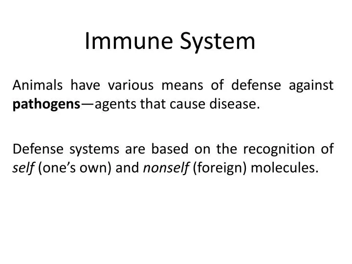
Immune System. Animals have various means of defense against pathogens —agents that cause disease. Defense systems are based on the recognition of self (one’s own) and nonself (foreign) molecules. Two general types of defense mechanisms:

E N D
Immune System Animals have various means of defense against pathogens—agents that cause disease. Defense systems are based on the recognition of self (one’s own) and nonself (foreign) molecules.
Two general types of defense mechanisms: Nonspecific defenses: protect against anything & everything (aka Innate) Specific defenses: aimed at specific pathogens (aka Adaptive)
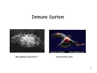
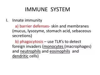
Non-Specific Immunity Barriers: skin mucous membranes cilia & mucus normal flora phagocytosis by macrophages inflammation
Blood plasma + RBCs + WBCs + platelets transport for both non-specific and specific Lymph derived from blood; returns fluid from tissue to circulation nodes- cells of the immune system reside here and check fluid for pathogens
Red and white blood cells originate from pluripotent stem cells in the bone marrow. These cells constantly divide and can differentiate into a variety of blood cells.
Specific Immunity Antibodies are proteins that bind to substances identified as nonself. Secreted by B cells. T cell receptors are integral membrane proteins, recognize and bind nonself molecules on other cells. Major histocompatibility complex (MHC): on the surface of most mammalian cells. They are self-identifying labels. Antigens: protein or part of protein-flag = not me
T cell receptors and antibodies bind to specific nonself molecules (antigens). Specific sites on the antigens are called antigenic determinants or epitopes.
The specific immune system has four key traits: • Specificity • Diversity—response to a wide variety of pathogens • Ability to distinguish self from nonself • Memory
Humoral vs. Cellular Response Antibody reacts to Antigen in blood, lymph & tissue fluids Triggers B cells to clone itself plasma cells make Antibody that binds to pathogen memory cells-perpetuate clone & reduce response time at 2nd exposure T cell receptor recognizes Antigen + MHC (me signal) Triggers T cells to clone itself T-cytotoxic –destroys cells with MHC1 + antigen T-helper-alerted by MHC2 + antigen-stimulates B cells to proliferate T-suppressor-turn off response
Figure 24.7A 1 2 3 4 5 6 Primary immune response Secondary immune response Antigenmolecules Antibodymolecules Antigen receptoron the cellsurface B cells withdifferentantigenreceptors First exposureto the antigen Cell activation:growth, division,and differentiation Clone of plasma (effector) cellssecreting antibodies Antigenmolecules Second clone Antibodymolecules Second exposureto the same antigen First clone Endoplasmicreticulum Clone of memory cells Memory cells Plasma (effector) cells secreting antibodies
Figure 24.7A_s1 1 Primary immune response Antigen receptoron the cellsurface B cells withdifferentantigenreceptors
Figure 24.7A_s2 1 2 Primary immune response Antigenmolecules Antigen receptoron the cellsurface B cells withdifferentantigenreceptors
Figure 24.7A_s3 1 2 3 Primary immune response Antigenmolecules Antigen receptoron the cellsurface B cells withdifferentantigenreceptors First exposureto the antigen Cell activation:growth, division,and differentiation
Figure 24.7A_s4 1 2 3 4 5 Primary immune response Antigenmolecules Antigen receptoron the cellsurface B cells withdifferentantigenreceptors First exposureto the antigen Cell activation:growth, division,and differentiation Antibodymolecules First clone Endoplasmicreticulum Plasma (effector) cells secreting antibodies Memory cells
Figure 24.7A_s5 6 Antigenmolecules Second exposureto the same antigen Memory cells
Figure 24.7A_s6 6 Secondary immune response Antibodymolecules Antigenmolecules Clone of plasma (effector) cellssecreting antibodies Second exposureto the same antigen Second clone Clone of memory cells Memory cells
Figure 24.7B Second exposureto antigen X,first exposureto antigen Y Secondary immuneresponse toantigen X First exposure to antigen X Antibody concentration Primary immuneresponse toantigen X Primary immuneresponse toantigen Y Antibodiesto Y Antibodiesto X 56 0 14 35 42 7 21 28 49 Time (days)
Figure 24.9 Binding of antibodies to antigensinactivates antigens by Neutralization(blocks viral binding sites;coats bacteria) Precipitation ofdissolved antigens Activation of thecomplement system Agglutinationof microbes Complementmolecule Bacteria Virus Antigenmolecules Bacterium Foreign cell Hole Leads to Enhances Phagocytosis Cell lysis Macrophage
Humoral vs. Cellular Response Antibody reacts to Antigen in blood, lymph & tissue fluids Triggers B cells to clone itself plasma cells make Antibody that binds to pathogen memory cells-perpetuate clone & reduce response time at 2nd exposure T cell receptor recognizes Antigen + MHC (me signal) Triggers T cells to clone itself T-cytotoxic –destroys cells with MHC1 + antigen T-helper-alerted by MHC2 + antigen-stimulates B cells to proliferate T-suppressor-turn off response
Figure 24.11 6 5 4 3 2 7 1 Humoralimmune response(secretion ofantibodies byplasma cells) Phagocytic cell(yellow) engulfinga foreign cell B cell Self-nonselfcomplex Interleukin-2stimulatescell division T cellreceptor Macrophage Microbe Interleukin-2activates B cellsand other T cells HelperT cell Cell-mediatedimmune response(attack oninfected cells) Self protein Bindingsite for theself protein CytotoxicT cell Antigen-presentingcell Interleukin-1stimulates thehelper T cell Antigen from the microbe(nonself molecule) Bindingsite for theantigen
Figure 24.11_1 3 2 1 Self-nonselfcomplex Macrophage Microbe Self protein Antigen-presentingcell Antigen from the microbe(nonself molecule)
Figure 24.11_2 3 2 4 5 6 7 Self-nonselfcomplex B cell Interleukin-2stimulatescell division T cellreceptor Interleukin-2activates B cellsand other T cells HelperT cell Bindingsite for theself protein CytotoxicT cell Antigen-presentingcell Interleukin-1stimulates thehelper T cell Bindingsite for theantigen
Figure 24.12_s1 1 A cytotoxic T cell bindsto an infected cell. Self-nonselfcomplex Foreignantigen Infected cell CytotoxicT cell Perforinmolecule
Figure 24.12_s2 2 1 Perforin makes holes in theinfected cell’s membrane,and an enzyme thatpromotes apoptosis enters. A cytotoxic T cell bindsto an infected cell. Self-nonselfcomplex A hole forming Foreignantigen Infected cell Enzymes thatpromote apoptosis CytotoxicT cell Perforinmolecule
Figure 24.12_s3 3 2 1 Perforin makes holes in theinfected cell’s membrane,and an enzyme thatpromotes apoptosis enters. The infected cellis destroyed. A cytotoxic T cell bindsto an infected cell. Self-nonselfcomplex A hole forming Foreignantigen Infected cell Enzymes thatpromote apoptosis CytotoxicT cell Perforinmolecule
Problems • Allergy-hypersensitive immune repsonse • stimulates release of histamine b/c of antigen-antibody interaction (immediate response) • T cell response initiated (delayed response) • Autoimmune • fail to destroy antibody producing cell that matches to self antigens • virus antigen resembles self antigen • T cells recognize antigen (non-self) that has a portion similar to self antigen
Figure 24.17 2 5 3 4 1 Sensitization: Initial exposure to an allergen Later exposure to the same allergen B cell(plasma cell) Mastcell Antigenic determinant Histamine B cells makeantibodies. The allergen bindsto antibodies ona mast cell. Antibodiesattach to amast cell. Histamine isreleased, causingallergy symptoms. An allergen (pollengrain) enters thebloodstream.