Download
1 / 33
520 likes | 1.92k Vues
Myocardial Infarction. By Dr. Hanan Said Ali. Learning Objectives. Define of myocardial infarction. Explain Pathophysiology of MI. Enumerate Clinical Manifestation of MI. Identify the diagnostic measures of MI. Discus complication of myocardial infarction.

E N D
Myocardial Infarction By Dr. Hanan Said Ali
Learning Objectives Define of myocardial infarction. Explain Pathophysiology of MI. Enumerate Clinical Manifestation of MI. Identify the diagnostic measures of MI. Discus complication of myocardial infarction. Explain the management and health teaching for client with MI .
What is 'acute coronary syndrome'? • The term 'acute coronary syndrome' is a term that is used more and more by doctors. It covers a range of disorders (including MI) that are caused by the same underlying problem. • The underlying problem is a sudden reduction of blood flow to part of the heart muscle. This is caused by a blood clot that forms on a patch of atheroma within a coronary artery (which is described earlier).
What is 'acute coronary syndrome'? • If the blood clot causes a reduced blood flow, but not a total blockage then the heart muscle supplied by the affected artery does not infarct (die). • This situation causes 'acute coronary syndrome with unstable angina' - and typically leads to a sudden worsening of angina pains.
What is 'acute coronary syndrome'? • If there is death of heart tissue then this is called an 'acute coronary syndrome with MI' (the subject of this leaflet). • There is a third 'in between' category where just a very small amount of heart tissue infarcts. This is called 'acute coronary syndrome with myocyte necrosis'. In effect, this is like having a mild MI.
What is 'acute coronary syndrome'? • One test that is used to distinguish between these three acute coronary syndromes is the blood test for troponin. • If the level of troponin is normal, then there is no death of heart tissue. • If the level is high, then it is classed as an MI. • If there is just a slight rise in the level of troponin then this diagnoses 'acute coronary syndrome with myocyte necrosis'.
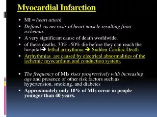
Definition of Myocardial Infarction • Myocardial infarction occurs when ischemic intracellular changes become irreversible and necrosis results.
Pathophysiology • Cardiac cells can withstand ischemia condition for approximately 20 minute before cellular death (necrosis)begins. • Contractile function of the heart stops in the area of myocardial necrosis. • A transmular MI occurs when the entire thickness of the myocardium in a region involved
Pathophysiology Cont. • A subendocardial MI (non- transmular) exists when the damage has not penetrated through the entire thickness of the myocardial wall. A myocardial infarction occurs when an atheroscleroticplaqueslowly builds up in the inner lining of a coronary artery and then suddenly ruptures, totally occluding the artery and preventing blood flow downstream.
Pathophysiology Cont. • If impaired blood flow to the heart lasts long enough, the heart cells die (chiefly through necrosis) and do not grow back. A collagenscar forms in its place. • This scar tissue also puts the patient at risk for potentially life threatening arrhythmias, and may result in the formation of a ventricular aneurysm that can rupture with catastrophic consequences.
Pathophysiology Cont. Posteroinferior infarct Anterolateral infarct
Clinical Manifestation • Chest pain is severe, diffuse steady substernal pain of a crushing and squeezing nature; not relieved by rest or sublingual vasodilator therapy, but requires narcotics; may radiate to the arms (commonly the left), shoulders, neck, back, and/or jaw; continues for more than 15 minutes
Clinical Manifestation Cont. • Nausea and vomiting As a result of vasovagal reflexes initiated from the area of the infarcted myocardium. • Sympathetic nervous system stimulation IS increased due to release of increased catecholemine (norepinephrine)and epinephrine leads to cool and clammy skin (cold sweats)
Clinical Manifestation Cont. • Fever -The temperature may increase up to 38c • Cardiac vascular manifestation - Includes elevated BP and HR. Later, BP may drop as decreased cardiac output, urinary output may be decreased. - Crackles. - later hepatic enlargement - Peripheral oedema indicate cardiac failure.
Diagnostic Measures • The client history of chest pain. • 12-Lead ECG. Consistent with acute MI.(ST-T wave elevated by greater than 1mm or more in two continuous leads . • Myocardial serum enzyme. • Echocardiogram. • Cardiac Imaging.
Complication of myocardial Infarction: 1. Arrhythmias: • The most common complications after MI are arrhythmias, resent in 80% of MI patients. Arrhythmias are caused by any condition that affects the myocardial sensitivity to nerve impulses, such as ischemia, electrolyte imbalances, and sympathetic nervous system stimulation
Complication of myocardial Infarction: 2. Right ventricular infarction: • Infarctions that primarily cause damage to the right ventricle (RV) are often seen with large inferior, inferolateral, or inferoposterior MIS. 4. Pericarditis: • Acute pericarditis, an inflammation of the visceral or parietal pericardium, or both, may result in cardiac compression, decreased ventricular filling and emptying, and cardiac failure. It may occur 2 to 3 days after an acute MI as a common complication of the infarction.
Complication of myocardial Infarction: 3. Ventricular aneurysms: • They are serious complications of transmular myocardial infarction, leading to severe hemodynamic compromise (heart failure, thrombo -embolism angina and arrhythmias). • It results when the infarcted, myocardial becomes thinned and bulges out during contraction. Surgery has been frequently indicated, and it improves symptoms and the quality of life.
Complication of myocardial Infarction: 5. Congestive heart failure (CHF): • It is a complication that occurs when the pumping power of the heart has diminished. In the patient with an acute MI it is common to see some degree of LV dysfunction in the first 24 hours. 6. Pulmonary embolism: • It may be seen in the patient with MI who has had bouts of CHF or has been extremely immobile because of prolonged bed rest.
Complication of myocardial Infarction: 7. Dressler's syndrome (post-MI syndrome): • It is characterized by pericarditis with effusion and fever that develops 1 to 4 weeks after MI. It may also occur after open-heart surgery. It is thought to be caused by an antigen-antibody reaction to the necrotic myocardium. 8. Cardiogenic shock: • It occurs when inadequate oxygen and nutrients are supplied to the tissues because of sever L.V failure. It occurs when there is loss of function of at least 40% of the LV. because of infarction.
Management Cont. In initial stages, of emergency management ,the nursing responsibility: • Ensure patent airway. • Insert two IV catheters. • Obtain 12-lead ECG. • Determine location of pain- Assess severity using pain , scale (0-10). • Medicate for pain as ordered. • Obtain cardiac enzyme levels.
Management Cont. Ongoing monitoring by nurses include : • Monitor vital sign , level of consciousness, cardiac rhythm , and O2 saturation, pain . • Anticipate need for intubation if respiratory distress is evident. • Prepare for CPR, defibrillation, transcutaneous pacing .
Management Cont. Common drugs used for coronary artery disease 1. Antiplatelet agents(asprin) • Inhibits platelet aggregation. 2. Nitrates. 3. Beta blocker. 4. Calcium channel blockers. 5. Heparin • Prevents propagations of established thrombus by rapidly inhibiting thrombin. PTTs /6 hours
Management Cont. 6. Thrombolytic (Streptokinase. to activate plasmin for lysis of obstructive Clots specific, therefore , systemic lysis may occur The nurse monitors for reperfusion , and bleeding. 7. Morphine sulphate Blunt the deleterious consequences of sympathetic stimulation with pain , and vasodilator monitor..... Hypotension, vital signs, level of consc. Dose ..............2-5 mg
Management Cont. 8. Cholesterol lowering agent. • They reduce the substance for lipid deposition in the coronary artery. • Lipid levels should be obtained at regular intervals. • Client must educated that cholesterol- lowering agents do not substitute for dietary modification. 9. Angiotensin- converting enzyme inhibitor
Management Cont. Coronary artery diseases may be treated with: • Intr-aortic balloon(IABP) • Percutaneous transluminal coronary angioplasty(PTCA) • Intracoronary stunting. • Coronary artery bypass graft.
Management Cont. Health Teaching: • Use and storage of nitroglycerin. • Risk factors modification. • Resumption of activities