Download

1 / 43
510 likes | 888 Vues
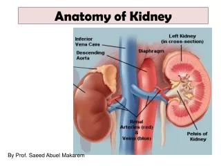
Pathology of Kidney. Dr. Sachin Kale, MD. Associate Professor, Dept of Pathology. Anatomy of Kidney. Note the positions of Glomerulus PCT, DCT, CT Cortex, Medulla, Pelvis. Glomerular diseases:. Primary Acute diffuse post streptococcal RPGN Membranous GN FSGS MPGN

E N D
Pathology of Kidney Dr. Sachin Kale, MD. Associate Professor, Dept of Pathology.
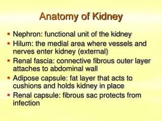
Anatomy of Kidney Note the positions of Glomerulus PCT, DCT, CT Cortex, Medulla, Pelvis.
Glomerular diseases: • Primary • Acute diffuse post streptococcal • RPGN • Membranous GN • FSGS • MPGN • Lipoid nephrosis or minimal change • IgA nephropathy • Secondary • SLE, Diabetes, Amyloidosis, Goodpasture’s syndrome, PAN, WG, HSP, Hypertension etc.
Clinical Syndromes: • Nephritic syndrome. • Oliguria, Haematuria, Proteinuria, Oedema, Azotemmia, Hypertension. • Nephrotic syndrome. • >3.5 gm proteinuria, Hypoalbuminemia hyperlipidemia, Lipiduria • RPGN. • Nephritis, loss of Kidney function - within weeks • Chronic renal failure. • Azotemia/uremia progressing over months and years • Asymptomatic Hematuria or proteinuria
CHRONIC RENAL FAILURE Fluid and Electrolytes:Dehydration, Edema, Hyperkalemia, Metabolicacidosis Calcium Phosphate and Bone:Hyperphosphatemia, Hypocalcemia, Secondary hyperparathyroidism, Renal osteodystrophy Hematologic:Anemia, Bleeding diathesis Cardiopulmonary:Hypertension, Congestive heart failure, Pulmonary edema, Uremic pericarditis Gastrointestinal:Nausea and vomiting, Bleeding, Esophagitis, gastritis, colitis Neuromuscular:Myopathy, Peripheral neuropathy, Encephalopathy Dermatologic:Sallow (greenish-yellow) color, Pruritus, Dermatitis
ACUTE TUBULAR NECROSIS • Destruction of renal TUBULAR epithelium • Loss of renal function • 50% of ACUTE renal failure • Two types: ISCHEMIC NEPHROTOXIC -AMINOGLYCOSIDES -AMPHOTERICIN B -CONTRAST AGENTS
ATN PATHOGENESIS • BLOOD FLOW DISTURBANCES (ISCHEMIC) • TUBULAR INJURY (NEPHROTOXIC)
CLINICAL COURSE • INITIATION (36 hours) • Mild OLIGURIA • Mild AZOTEMIA • MAINTENANCE • More OLIGURIA • More AZOTEMIA • DIALYSIS NEEDED • RECOVERY • HYPOKALEMIA main problem • BUN, CREATININE return to normal
Immune Mechanisms of Glomerular injury: • Antibody mediated: • In-Situ immune complex deposition • Tissue antigens - Goodpasture anti GBM Ag • Planted antigens - infections, toxins, drugs. • Circulating immune complex deposition. • Endogenous - DNA as in SLE • Exogenous – infections – HBsAg, Syphilis, Streptococcal, Falciparum, • Cell mediated Immune injury • Activation of alternate complement pathway
Immune Glomerulonephritis: • Antigen or Antibody - Immune reaction • Activation of complements, Neutrophils… • destruction of glomerular structure • Inflammation, exudation swelling. • ↓ blood flow, GFR, - • Oliguria, Proteinuria, Hematuria, Hypertension.
Neutrophil Activity • Proteases – GBM degradation • Reactive oxygen metabolites – cell damage • Arachidonic acid metabolites –Reduction in GFR
Other Mediators • Cytotoxic antibodies • Macrophages • Platelets • Resident glomerular cells • Fibrin related products
Nephritic Syndromes : • Diffuse Proliferative GN • Post Streptococcal. • Rapidly Progressive GN (or Crescentic) • Post Streptococcal, Goodpasture’s, • Focal Glomerulonephritis • Primary: Bergers disease (IgA Nephritis) • Secondary IgA nephritis, Henoch Schonlein purpura, SBE, Coeliac Disease etc.
Diffuse Proliferative GN: • Post streptococcal* common – • Primary infection - Pharynx, skin, ear etc.. • Kidney damage – 1-4 weeks after infection. • Malaise, fever, nausea, edema*, ↑ASO, ↓C3 • Resolution in 6-8 weeks.
Post Streptococcal GN (Prol.GN): • 1-4 weeks following streptococcal infection by nephritogenic strains (time for Ab formation) • Immune mediated • Granular deposits of IgG,IgM & C3 in GBM, (subepithelial location common) • Humps in GBM on EM or IF Microscopy
Normal • Inflammation • Proliferation • Swelling. • Narrow capillary • ↓GFR-Renin-BP • Post Strepto GN
Diffuse Proliferative GN: • Enlarged hypercellular glomeruli. • Hyperplasia of epithelium & endothelium. Cell Swelling. • Inflammatory cells. • Collapsed capillaries. Obstruction to blood flow.
Pathogenesis of Diffuse PGN: • Streptococcal infection – Antibody attack GBM - inflammation & proliferation. • Glomerular capillary obstruction: • J.G.A stimulation – Renin – high blood pressure • Reduced filtration – raised blood urea • Fluid retention – Oedema • Damage to GBM: • Unselective proteinuria (form Pr. casts in tubule) • Haematuria (form RBC casts in tubule)
Progression of DPGN: Rapidly Progressive GN Poststreptococcal DPGN Complete Healing Cardiac Failure or Uremia; death in acute phase CGN
RPGN • Clinicopathologic syndrome • Glomerular damage • Rapid progressive decline in renal function • Histology: accumulation of cells in Bowman’s space in the form of “Crescents”
RPGN: Classification & Pathogenesis • Postinfectious • GN associated with systemic diseases • Idiopathic RPGN • Glomerular injury is immunologically mediated. • Goodpasture’s syndrome – classic anti-GBM nephritis
RPGN classification • Post-infectious RPGN • Systemic diseases – • SLE, Goodpasture’s, Vasculitis (PAN), Wegener’s granulomatosus, HSP, Essential cryoglobulinemia • Idiopathic RPGN
RPGN cont.. • Idiopathic : ½ the cases, • Linear, Granular or minimal to none immune deposits • Gross: Enlarged pale kidneys Large white kidney • Petechial hemorrhages in cortex • M/E: Glomeruli: focal necrosis, endothelial proliferation
RPGN… • Formation of crescents: • Proliferation of parietal cells, migration of monocytes and macrophages into Bowman’s space • Crescents obliterate Bowman’s space, compression capillary tuft • Crescents undergo sclerosis
RPGN: Clinical features • Goodpasture’s Syndrome: recurrent hemoptasis & renal manifestations • Hematuria, Red cell casts, Moderate proteinuria, • Variable HT and edema • Oliguria
Which of the following presents with hematuria, proteinuria and hypertension • Nephrotic syndrome • Nephritic Syndrome • UTI • Renal Tubular Acidosis
All of the following are seen in renal failure except • Hypercalcemia • Hyperkalemia • Bone lesions • Metobolic Acidosis
Anemia in renal failure is generally • Microcytic hypochromic • Normocytic normochromic • Dimorphic • megaloblastic
Which of the following is not a primary GN • Minimal Change disease • Membranous GN • Diabetes mellitus • RPGN
Which of the following is not part of nephrotic syndrome • Lipiduria • Hypertension • Proteinuria • Edema
True about Post-strepto GN - • Occurs 1 – 4 months after infection • Occurs 1 – 4 days after infection • Occurs 1 – 4 weeks after infection • Non of the above
False about RPGN.. • Formation of crescents • Small contracted kidneys • Hematuria • Oliguria
Spot the diagnosis RPGN
Spot the diagnosis Post streptococcal GN
Thought for the day… • Ours is a world where people don't know what they want and are willing to go through hell to get it.
Thanks… • http://sachinkale1.tripod.com