Download
1 / 19
430 likes | 1.24k Vues
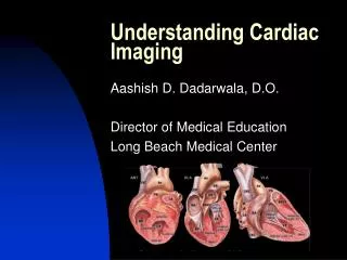
BY NICOLE BROOKER. Understanding Cardiac Electrophysiology. TABLE OF CONTENTS. Electrical conduction system Electrical conduction pathway Conduction components: The sinoatrial node (SA) Bachmann’s bundle Internodal tracts

E N D
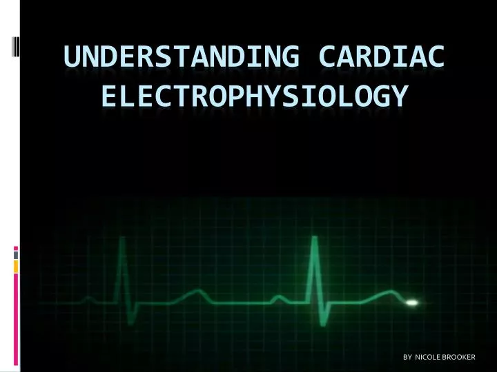
BY NICOLE BROOKER Understanding Cardiac Electrophysiology
TABLE OF CONTENTS • Electrical conduction system • Electrical conduction pathway • Conduction components: The sinoatrial node (SA) Bachmann’s bundle Internodal tracts Atrioventricular node (AV) Right and Left Bundle branches Perkinje fibers Ventricles • Basic electrocardiograph (EKG) waveform: P wave PR interval QRS complex ST segment T wave QT interval
Electrical Conduction System of the Heart • The heart is supplied with an electrical conduction system that generates and conducts electrical impulses along specialized pathways • Contraction is sequence specific so the atria contracts before the ventricle and the ventricle contracts from apex to base for efficient ejection of blood.
CONDUCTION COMPONENTS • Sinoatrial (SA) node • Interatrial tract (Bachmann’s bundle) • Internodal tracts, the atrioventricular (AV) node • Bundle of His • Right and left bundle branches • Purkinje fibers.
THE SINOATRIAL NODE (SA) • The SA node, located in the upper right atrium, is the primary pacemaker of the heart (60 to 100 beats). • If the SA node fails to generate impulses or if those impulses are blocked, pacemaker cells in other sites can assume control, but at a slower rate.
BACHMANN’S BUNDLEINTERNODAL TRACTS • Bachmann’s bundle conducts impulses to the left atrium, while the internodal tracts conduct impulses to the AV node, located in the lower R atrium near the interatrial septum.
ATRIOVENTRICULAR NODE • Primary function-slow conduction of electrical impulses coming from the atria, allowing time for the atria to contract and empty their contents into the ventricles. • It can functions as a backup pacemaker (40 to 60 beats). When the atrial rate is rapid, the AV node blocks some of the impulses being conducted to the ventricles, protecting them from dangerously fast rates.
RT AND LT BUNDLE BRANCHES • From the AV node, the electrical impulse moves rapidly through the bundle of His and to the R bundle branch (R ventricle) and L bundle branch ( L ventricle).
PURKINJE FIBERS • The impulse then enters the Purkinje system where Purkinje fibers conduct the impulse to myocardial cells of the ventricle, causing ventricular depolarization and contraction.
THE VENTRICLES • The ventricles also have pacemaker cells (30 to 40 beats) that take over if impulses are not being transmitted by the SA or AV nodes. • Repolarization follows.
Basic ECG Waveform • The heart’s electrical activity is represented by an ECG tracing by three basic waveforms: the P wave, the QRS complex, and the T wave.
BASIC ECG WAVEFORM • Between the waveforms are the following segments and intervals: the PR interval, the ST segment, and the QT interval. A U wave is sometimes present.
THE P WAVE • P wave - atrial depolarization, or the spread of the impulse from the SA node throughout the atria.
THE PR INTERVAL • PR interval - the time required for the impulse to leave the SA node, travel through the atria, AV node, bundle of His, bundle branches, and Purkinje fibers.
THE QRS COMPLEX • QRS Complex-represents ventricular depolarization and is represented by three waves: Q, R and S.
THE T WAVE • T wave - the latter phase of ventricular repolarization, and the vulnerable period of repolarization (R on T phenomenon).
THE ST SEGMENT • ST segment - represents the end of ventricular depolarization and the beginning of ventricular repolarization.
THE QT INTERVAL • QT interval - the time between the onset of ventricular depolarization and the end of ventricular repolarization • Includes the QRS complex, ST segment, and T wave