Download
1 / 23
240 likes | 379 Vues
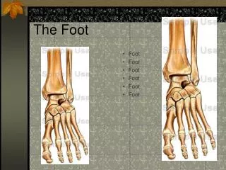
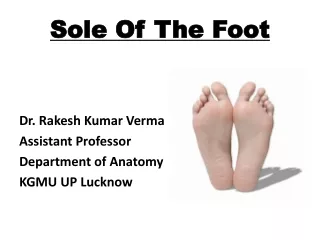
Anatomy of the Foot. Bones Joints Muscles Skin. Bones of the Foot. Basic Facts: There are 26 bones in each human foot. Makes up one quarter of the entire (208 bones) human body. 2 in the hind foot or rear foot 5 in the midfoot 19 in the forefoot

E N D
Anatomy of the Foot Bones Joints Muscles Skin
Bones of the Foot Basic Facts: There are 26 bones in each human foot. Makes up one quarter of the entire (208 bones) human body. 2 in the hind foot or rear foot 5 in the midfoot 19 in the forefoot 2 ancillary bones underneath the first metatarsal head.
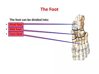
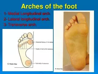
Medial View of Foot Midfoot Navicular, cuneiforms (3) and cuboid Rear foot/Hind foot Talus and calcaneus Forefoot Metatarsals (5) and phalanges (14)
Lateral View of the foot Midfoot Forefoot Hind foot/Rear foot
Dorsal view The 5 metatarsal bones are made up of three main parts—the base, the shaft and the head. The base is at the proximal end. The shaft is in the middle. The heads are located at the distal ends of the bones. The heads are the weight bearing portion of the foot. The metatarsals are numbered 1-5 beginning with the great toe, or hallux.
Sesamoids Located on the plantar side of the great toe (hallux). Identified by the location on the foot-- tibial (medial) and fibular (lateral) sesamoids. Flexor hallucis longus tendon runs between them. The tendon is responsible for abducting and adducting great toe.
Ankle Bones • Posterior view of ankle • Tibia • Medial malleolus (tibia) • Lateral malleolus (fibula) • Talus (slightly displaced) • Calcaneus 5
Circulatory System • There are three types of blood vessels: • Arteries-carries blood away from the heart • Veins-transfers blood back to the heart from the capillaries • Capillaries-connect arteries to veins
Posterior Tibial Artery The posterior tibial artery is found on the medial side of the ankle and can be palpated.
DorsalisPedis Artery The dorsalis pedis artery can be palpated on the dorsum of the foot between the first and second ray.
Skin The human skin is the bodies largest organ and made up of multiple layers. The top layer of skin, called the epidermis, plays a very important role in protecting the body against the environment. Other functions include insulation, temperature regulation and sensation.
Skin Skin is divided into 3 main layers: Epidermis-outermost layer; visible Dermis-layer underneath epidermis Hypodermis- also called the subcutaneous tissue, used for fat storage. The thickness of the skin at the soles (and palms) is 1.5 mm at the epidermis layer.
Joints A joint is the area where two or more bones are attached for the purpose of motion of human body parts. A joint is usually formed of fibrous connective tissue and cartilage. There are 33 joints in the human foot. They allow the foot to move in many directions and adapt to many surfaces. The joints are shock absorbers.
Joints • The metarso-phalangeal joint (MTP) is between the metatarsals and the phalanges (toes). • Hinge joints that allow mostly plantar and dorsiflexion and also allow the toes to maintain contact with the ground during push off.
Joints • The proximal interphalangeal joint (PIP) is between the proximal and middle phalanges. • The distal interphalangeal joint (DIP) is found only on phalanges 2-5.
Joints • The ankle joint is composed of the fibula, tibia and talus. • A hinge joint that allows the foot to pull up (dorsiflex) and move downward (plantarflex). Anterior view Posterior view
Joints • The transverse tarsal joint is comprised of two joints—the talonavicular and calcaneocuboid (TNCC joint). • Also known as the midtarsal or chopart’s joint
Joints • The tarsometatarsal joint is made up of the tarsals and metatarsals. • Also known as the Lisfranc Joint.
Joints • The subtalar joint is between the talus and calcaneus. • Also known as the talocalcaneal joint. • Acts as a screw-shaped joint and is the primary joint that allows the foot to turn in (inversion) or turn outward (eversion) Posterior view
Bursae • Bursae, or singular bursa, fluid filled sacs commonly located just proximal to insertion of tendon into bone; reduce friction that would otherwise occur as tendon moves against bone
Muscles and Tendons Muscles are attached to tendons which insert to the bones of the foot and toes. Most muscles that move the foot originate from the calf (posterior) or shin (anterior) area. Tendons are a band of fibrous tissue that attach muscles to the bones. When a muscle contracts, it pulls on the tendon. One of the most prominent tendons is the Achilles tendon, which can be felt on the back of the foot, just above the heel. Tendons can be torn or ruptured and inflammation is the most prevalent.
Muscles • The gastrocenimus and soleus muscle combine to form the Achilles tendon. • This allows the ankle and foot to push down (plantar flex).
Achilles tendon • Most important tendon for walking, running and jumping. • Attached the calf muscle to the calcaneus. • Allows us to plantar flex. • The strongest and thickest tendon.