Download
1 / 56
560 likes | 668 Vues
Biology 221 Anatomy & Physiology II. TOPIC 6 Immune System Resistance to Disease. Chapter 21 pp. 778-787. E. Lathrop-Davis / E. Gorski / S. Kabrhel. Overview: Functions. Functional rather than anatomical system Functions : Protects against pathogens microbes parasites

E N D
Biology 221 Anatomy & Physiology II TOPIC 6 Immune SystemResistance to Disease Chapter 21 pp. 778-787 E. Lathrop-Davis / E. Gorski / S. Kabrhel
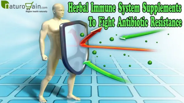
Overview: Functions • Functional rather than anatomical system • Functions: • Protects against pathogens • microbes • parasites • Eliminates tissues and cells that have been damaged, infected or killed • Distinguishes between self and non-self
Overview Two types of resistance work together against disease • Innate = nonspecific • general defense against wide range of pathogens • rapid response • in place at birth • mechanisms: intact membranes, phagocytes, antimicrobial chemicals, inflammation • Adaptive = specific • specific response to pathogens • slower than innate system • acquired as person is exposed • mechanisms: T cell lymphocytes, antibodies
Nonspecific Resistance - Overview • A. Physical barriers • B. Cellular barriers • C. Fever See Table 22.2, p. 801
Physical Barriers: Intact Skin • Consists of keratinized stratified squamous epithelium • Relatively dry (inhibits growth of some pathogens) • Sebaceous gland secretions include antibacterial chemicals (lysozyme, fatty acids) • Normal bacterial flora compete with pathogens • Slightly acidic • Slightly salty (sweat) Fig. 5.3, p. 152
Physical Barriers: Intact Mucous Membranes • Line body cavities open to outside (digestive, urinary, reproductive, respiratory tracts) • Intact barrier – nonkeratinized stratified squamous epithelium lines openings (mouth, pharynx, esophagus, vagina, parts of rectum and urethra) • Low pH in some areas • slightly acidic (mouth, vagina, urethra) to highly acidic (stomach) • Antimicrobial proteins (lysozyme in saliva and lacrimal fluid) • Normal bacterial flora compete with pathogens
Physical Barriers: Mucus • hairs help trap particles • cilia move particles • ciliary escalator -- present in trachea and bronchi; moves mucus and particles up to throat
Cellular Responses: Inflammation • Functions: • prevents spread of pathogens or damaging chemicals to other tissues • removes dead cells and pathogens • prepares tissue for repair • Signs of inflammation • redness, heat, swelling, pain
Main Inflammatory Chemicals • Histamine • secreted by basophils and mast cells • vasodilation and increased capillary permeability • antihistamines • Kinins (proteins, e.g., bradykinin) • vasodilation, increased capillary permeability • induce chemotaxis (draw WBCs to area) • stimulate pain receptors
Other Inflammatory Chemicals • Prostaglandins (lipid – fatty acid molecules) • sensitize blood vessels to other inflammatory chemicals (enhance inflammation) • stimulate pain receptors • Complement – enhances inflammation • Cytokines • proteins released by various WBCs and tissue cells • many enhance various aspects of inflammation
Process of Inflammation • Release of inflammatory chemicals • Vascular changes: vasodilation and increased capillary permeability, resulting in: • hyperemia (increased blood flow to area) and exudate formation (loss of fluid containing plasma proteins to tissue) • increased temperature increased cellular metabolism See Fig. 22.2, p. 797
Process of Inflammation • increased oxygen and nutrients to tissue and cellular defenders • leakage of clotting proteins walls off pathogen to limit spread; forms network for tissue repair See Fig. 22.2, p. 797
Process of Inflammation (con’t) • Phagocyte mobilization • leukocytosis - increased number of leukocytes (especially neutrophils) • chemotaxis draws leukocytes to injured area • margination (“pavementing”; leukocytes adhere to capillary wall) • diapedesis (leukocytes pass through capillary wall) • phagocytosis of pathogens and debris • pus formation - occurs with severe infection (WBCs, dead and dying tissue cells, pathogens accumulate) See Fig. 22.3, p. 798
Phagocytes in Inflammation • Neutrophils respond most quickly (usually within a few hours) • associated with acute, local infection • Monocytes respond more slowly (usually within 8-12 hours) • enter tissue and become macrophages with more lysosomes • associated with chronic infection Think About It:What causes each of the signs of inflamamtion? - redness, heat, swelling, pain
Cellular Responses: Major Phagocytes • Macrophages • reside in tissues • derived from monocytes • free (wandering) macrophages - wander through tissues • fixed macrophages - stay in particular organ • e.g., Kuppfer cells (liver), microglia (brain) http://www.usc.edu/hsc/dental/ghisto/gi/c_90.html
Cellular Responses: Major Phagocytes • Neutrophils = microphages • respond quickly to localized infections • degranulation - release of chemicals stored in granules destroys pathogens also kills neutrophil http://www.usc.edu/hsc/dental/ghisto/bld/c_1.html
Cellular Responses: Other Phagocytes • Eosinophils • respond most to parasitic worms (release chemicals to destroy worm) http://www.usc.edu/hsc/dental/ghisto/bld/c_3.html
Cellular Responses: Other Phagocytes • Mast cells • reside in tissues • release histamine during inflammation • less common • respond to variety of bacteria (phagocytic role uncertain) http://image.bloodline.net/stories/storyReader$1682
Mechanism of Phagocytosis Steps: • Microbial adherence • recognition of bacterium as “non-self” • more difficult with encapsulated bacteria • opsonization – enhanced phagocytosis due to presence of complement proteins and antibodies • Formation of pseudopodia and engulfment of particle • Union of phagocytic vesicle with lysosome • Digestion of particle • Exocytosis of indigestible material See Fig. 22.1, p. 795
Mechanism of Phagocytosis (con’t) • Respiratory burst • used against pathogens that resist lysosomal enzymes (e.g., tuberculosis bacteria) • stimulated by chemicals released by immune system • produces free radicals (e.g., NO) • Defensins – antimicrobial proteins produced by neutrophils
Cellular Responses: Natural Killer (NK) Cells • Large, granular lymphocytes • Responsible for immunological surveillance– respond to abnormal antigens • Kill cancer cells and virally-infected cells • Release perforins • produce channels in target cell membrane • cause nucleus to degrade • Produce other chemicals that enhance inflammation
Antimicrobial Proteins: Complement • Group of 20+ plasma proteins (circulate in inactive form) • Two pathways of activation: • classical pathway • linked to immune system • activation results from interaction of antigen-antibody complex with key complement proteins • alternative pathway – interactions of other complement proteins with polysaccharides on surface of certain microorganisms
Antimicrobial Proteins: Complement • Both pathways start cascade resulting in • enhanced actions of nonspecific and specific resistance mechanisms, including inflammation and opsonization • lysis of bacterial cells Fig. 22.5, p. 800
Antimicrobial Proteins: IFNs Interferons • group of related proteins secreted by body cells infected with virus • alpha () & beta () stimulate synthesis of PKR in nearby uninfected cells • PKR is a protein that blocks protein synthesis at ribosomes --> prevents viral replication • gamma stimulate (activate) macrophages and NK cells • produced artificially and used clinically to treat genital herpes (caused by herpes virus), also used in treatment of hepatitis C, and viral infections in organ transplant patients
Antimicrobial Proteins: Lysozyme • In tears & saliva • Kills unencapsulated bacteria
Fever • Increased body temperature in response to pathogens • Involves resetting of “thermostat” in hypothalamus • response to pyrogens secreted by leukocytes and macrophages in response to bacteria and other foreign particles • Mild fever • enhances activity of phagocytes and tissue repair • causes liver and spleen to sequester iron and zinc (needed by bacteria to multiply) • High fever (> 104 oF or 40 oC) - damages proteins of sick person
Specific (Adaptive) Resistance Also known as acquired resistance Characteristics: • antigen specific • systemic • differentiates between normal (self) antigens and foreign (non-self) antigens • has memory (faster response second time around)
Specific (Adaptive) Resistance Types • Humoral = antibody-mediated immunity • result of specific antibodies (proteins) present in blood • Cellular = cell-mediated immunity • result of specific group of cells = T cell lymphocytes
Antigens (Ags) • Substances that activate immune system and elicit response • immunogenicity - cause production of antibody by plasma cells • reactivity - reacts with antibody, if present • Antigenic determinants = epitopes • parts of antigen that are recognized by T cells and antibodies • usually protein- or sugar-based Fig. 22.6, p. 803
Antigens (con’t) • Complete antigen has both characteristics • large molecules typically with more than one antigenic determinant • most foreign proteins, nucleic acids, some lipids, some large polysaccharides • Haptens = incomplete antigens – reactive but not immunogenic • generally small molecules • hapten can combine with other molecules to become complete antigen • e.g., penicillin
Antigens (con’t) • Self-antigens – major histocompatibility complex (MHC) proteins • glycoproteins found on individual’s own cells • two types: • Class I MHC proteins – found on all cells of body • Class II MHC proteins – found only on cells involved in immune response
Antigens (Ags): Terms • Agglutination – antibody binds to antigenic determinants of cells and cross-links several together resulting in clumping • e.g., cross-reactions between blood types • Precipitation – antibody binds to antigenic determinants of soluble antigen (e.g., toxin) and causes clumping • Neutralization – antibody covers active site(s) on antigen
Cells of the Immune System Lymphocytes • become immunocompetent in primary lymphoid organs (bone marrow or thymus), where they learn self-tolerance (recognition of body’s own protein antigens) • move to secondary lymphoid tissue to become exposed to antigens, then return to blood and lymph circulation • types: • B cells = B lymphocytes • T cells = T lymphocytes • Antigen-presenting cells (APCs)
Cells of the Immune System • B cells = B lymphocytes • become immunocompetent in bone marrow • develop into plasma cells after exposure to antigen and produce specific antibodies • T cells = T lymphocytes • become immunocompetent in thymus • active in cellular immunity Fig. 22.8, p. 805
Cells of the Immune System: APCs Antigen-presenting cells (APCs) • types: • dendritic cells • Langerhan’s cells of epidermis • macrophages • activated B cell lymphocytes • engulf foreign particles and present fragments on own surface to T cells so that the latter can recognize and respond to them
Humoral Immunity • Relies on B cells • Primary response – to first exposure • antigen binds to B cell with appropriate receptor B cell engulfs antigen divides into daughter cells that secrete antibodies or become memory cells • Secondary response – much faster due to presence of memory B cells Fig. 22.9, p. 807
Passive Versus ActiveHumoral Immunity Active Passive Naturally Acquired Infection Antibodies passed from mother to fetus or infant Artificially Acquired Vaccine (dead or attenuated pathogens) Injection of gamma globulin
Antibodies • Immunoglobulins (Igs) = gamma globulins • General structure: (See Table 22.3, p. 811) • consist of 4 polypeptide chains held together by disulfide bonds = antibody monomer • each chain has a variable and a constant region Fig. 22.12, p. 810
Antibodies • variable region • give specificity to antibody • includes antigen-binding sites • constant region • includes stem region of heavy chains and proximal parts of both heavy and light chains • stem region determines actions and classes of antibodies Fig. 22.12, p. 810
Most Common Antibody Classes • IgG • most abundant and diverse plasma antibody in both primary and secondary responses • protects against circulating bacteria, viruses, toxins • activates complement • crosses placenta to protect fetus • IgM • actgs as antigen receptor on B cell membrane • important to primary response • causes agglutination and activates complement
Less Common Antibody Classes • IgA • found primarily in mucus and other secretions (e.g, saliva, sweat, intestinal juice, milk) • prevents attachment of antigens to epithelium • IgD • acts as antigen receptor • IgE • present in skin, gastrointestinal and respiratory tract mucosae, tonsils • binds to mast cells and basophils • increases during allergy and chronic parasitic infection of GI tract
Mechanisms of Antibody Action • Enhance phagocytosis by: • Neutralization • Agglutination • Precipitation • Activate complement,which: • Enhances inflammation • Causes cell lysis • Enhances phagocytosis Fig. 22.13, p. 812
Cell-Mediated Immunity • Involves T cells • Types of T cells (see Table 22.4, p. 818) • Cytotoxic T cells (TC) • Helper T cells (TH) • Suppressor T cells (TS) • Delayed hypersensitivity T cells (TDH)
Cytotoxic T Cells (TC) • Destroy body cells that are infected by antigen (viruses, bacteria, internal parasites) or have non-self antigens (e.g., cancer cells) • Mechanism seems to involve release of perforin onto membrane of affected cell Fig. 22.17, p. 820
Cytotoxic T Cells (TC) • Other mechanisms include: • lymphotoxin (causes fragmentation of target cell DNA) • tumor necrosis factor (TNF; triggers cell death = apoptosis) • gamma interferon (stimulates macrophages)
Helper T Cells (TH) • stimulate production of B cells and cytotoxic T cells Fig. 22.16, p. 817
Other Types of T Cells • Suppressor T cells (TS) – limit activity of T and B cells after infection has been beaten • Delayed hypersensitivity T cells (TDH) – • involved in delayed allergic reactions by secreting interferon and other cytokines • enhance nonspecific phagocytosis by macrophages
T Cell Activation • Step 1 – Antigen binding • T cell antigen receptor (TCR) binds to antigen-MHC protein complex on cell • Step 2 – Costimulation • recognition of costimulatory signals stimulates clonal division of T cells into various types • Cytokines • released by macrophages and T cells • some act as costimulators
Organ Transplants Types: • Autograft – from one site to another in same person (e.g., skin graft) • Isograft – between identical twins (or members of same clone) • Allograft – between nonidentical individuals of same species • Xenograft – between different species (e.g., pig valves in heart; baboon heart in infant)
Organ Transplant: Rejection • Occurs when antigens on donor tissue are attacked by recipient’s immune system • Immunosuppressive therapy • corticosteroids – suppress inflammation • cytotoxic drugs • radiation (X ray) therapy • antilymphocyte globulins • immunosuppressant drugs (e.g., cyclosporine)