Download
1 / 32
330 likes | 542 Vues
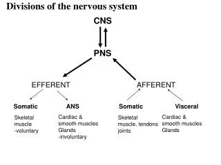
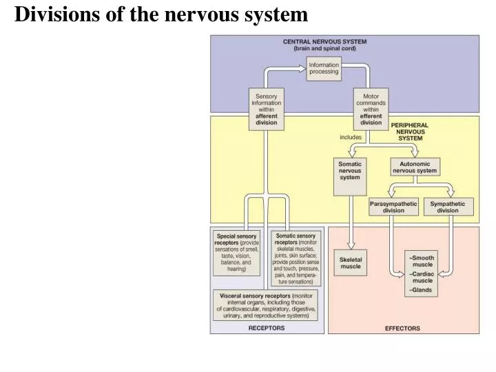
Divisions of the nervous system. PNS Terminology. Ganglia – neuron cell bodies Peripheral nerves – neuronal axons PNS neuroglia Satellite cells Enclose neuron cell bodies in ganglia Schwann cells Cover peripheral axons. Efferent Division of the PNS.

E N D
PNS Terminology • Ganglia – neuron cell bodies • Peripheral nerves – neuronal axons • PNS neuroglia • Satellite cells • Enclose neuron cell bodies in ganglia • Schwann cells • Cover peripheral axons
Efferent Division of the PNS • the somatic nervous system and part of the autonomic nervous system • the somatic – control of skeletal muscle • the ANS – involuntary control over cardiac and smooth muscle + gland secretion
I - Olfactory II - Optic III - Oculomotor IV-Trochlear V - Trigeminal VI - Abducens VII - Facial VIII - Acoustic IX - Glossopharyngeal X - Vagus XI - Accessory XII - Hypoglossal -cranial nerves – 12 pairs -considered part of the peripheral nervous system (PNS) -olfactory & optic contain only sensory axons = sensory nerves -remaining are motor or mixed nerves (both motor and sensory axons)
Spinal Nerve • after passing through intervertebral foramina the spinal nerve branches = ramus/rami • Dorsal ramus • Sensory/motor innervation to skin and muscles of back • Ventral ramus • -Ventrolateral body surface, body wall structures, muscles of the upper and lower limbs • in addition to these rami - the spinal nerves also give off a meningeal branch - reenters the vertebral canal and supplies the vertebrae, vertebral ligaments and meninges
sympathetic trunk • rami communicantes = branches from the spinal nerve • -defined as a connection between a spinal nerve and the sympathetic trunk of the ANS
Nerve Plexuses • Joining of ventral rami of spinal nerves to form nerve networks or plexuses • Found in neck, arm, low back & sacral regions • No plexus in thoracic region • intercostal nn. innervate intercostal spaces • T7 to T12 supply abdominal wall as well • Four major plexuses • Cervical plexus • Brachial plexus • Lumbar plexus • Sacral plexus
Somatic Nervous System • considered the voluntary aspect of the PNS • but the muscles of posture and balance are controlled involuntarily by the lower brain centers (brain stem, cerebellum) • cell bodies located in the ventral gray horn of the spinal cord • the axon of a motor neuron extends from the CNS continuously to its skeletal muscle target • ANS usually requires two neurons • terminals release acetylcholine – contraction • can only stimulate its target • whereas the ANS can either stimulate or inhibit its target
Somatic Nervous System - somatic/motor axons emerge from the ventral horn into the spinal nerve and travel through the: • dorsal ramus to target the muscles of the back • ventral ramus to target the muscles of the limbs and body wall (chest/abs/pelvis)
Somatic Nervous System • motor neurons receive incoming information from many converging presynaptic neurons • both excitatory and inhibitory on these motor neurons • some information are part of reflexes originating in the spinal cord • other information can come in from areas of the brain via the descending white matter tracts • motor areas of the cerebral cortex, the basal nuclei and the cerebellum • synapse with the motor neurons in the ventral horn and regulate their activity • activation – impulse sent to muscles • inhibition – no impulse, no contraction • level of activity of a motor neuron is the balance between the EPSPs/activation and the IPSPs/inhibition of the incoming synapsing neurons • therefore motor neurons are considered the final common pathway • considered the only way any other part of the nervous system can influence muscle activity
Somatic Motor pathways • all excitatory and inhibitory signals that control movement converge on the motor neurons that extend from the brain stem and SC to innervate the skeletal muscles • called lower motor neurons (LMNs) • have their cell bodies in the brain stem and SC • their axons extend through the cranial and spinal nerves to skeletal muscle • only LMNs provide output from the CNS to skeletal muscle fibers = final common pathway • damage to the LMNs produces flaccid paralysis on the same side as the damage – loss of reflex action, motor tone and voluntary contraction • neurons in four distinct circuits control movement by providing input to these LMNs • 1. local circuit neurons • 2. upper motor neurons (UMNs) • 3. basal ganglial neurons • 4. cerebellar neurons
Somatic Motor pathways • 1. local circuit • input arrives at LMNs from nearby interneurons called local circuit neurons • receive input from somatic sensory receptors and higher centers of the brain • help coordinate rhythmic activities in muscle groups • 2. UMNs • provide input to the local circuit and LMNs • essential for planning, initiating and directing sequences of voluntary movements • extend from the brain to the LMNs via two types of somatic motor pathways • 1. direct motor pathways: nerve impulses for voluntary movement • lateral corticospinal, anterior corticospinal and corticobulabar (brain stem) • about 90% decussate within the medulla oblongata (corticospinal) • originate in the cerebral cortex (motor cortex) and travel down the spinal cord (via white columns) to emerge as spinal nerves or through the brain stem and out as cranial nerves • 2. indirect motor pathways: or extrapyramidal pathways • nerve impulses follow complicated circuits that involve the cortex, basal ganglia, thalamus and brain stem
Somatic Motor pathways • 3. Basal ganglia • assist movement by providing input to the UMNs • also suppresses unwanted movements by inhibiting thalamus activity • the production of dopamine by the substantia nigra also effects muscle tone • caudate nucleus and putamen receive sensory input from several areas of the brain • output from the globus pallidus and substantia nigra goes to the motor areas of the cortex via the thalamus • this circuit (cortex – basal ganglia – thalamus – cortex) may function in initiating and terminating movements • 4. Cerebellar • function involves four activities • 1. monitoring intentions for movement • 2. monitoring actual movement • 3. comparing the command (intention and movement) with sensory information • 4. correction – to UMNs • travels via the thalamus to the UMNs in the cerebral cortex • or can go directly to the UMNs in the brain stem
The Neuromuscular Junction • end of neuron (synaptic terminal or axon bulb) in very close association • with a muscle fiber/cell • distance between the bulb and the folded sarcolemma = synaptic cleft • nerve impulse leads to release of neurotransmitter(acetylcholine) • this release will result in activation of the muscle cell and contraction • therefore the NMJ is ALWAYS excitatory • the only way inhibition can take place is through the inhibition of the neuron “connecting” with the muscle http://www.blackwellpublishing.com/matthews/neurotrans.html
ANS • two divisions that innervate the same organs • efferent branch regulates “visceral” activities (motor commands, involuntary, organs) • also has an afferent branch that receives sensory information from these areas
ANS • involuntary motor commands and sensory information • supplies cardiac and smooth muscle, glands (i.e. viscera) • comprised on two neurons • preganglionic and postganglionic • preganglionic synapses with the cell body of the postganglionic within the ganglion • the pregang and postgang neurotransmitters can differ • the postganglionic neuron is unmyelinated • glands are innervated by the preganglionic neuron – e.g adrenal gland which then releases epinephrine or norepinephrine in response
Parasympathetic Division • cell bodies of the preG neurons are located in the four cranial nerves III, VII, IX and X (brain stem) and in the lateral gray horns of the sacral spinal nerves 2 through 4 • emerge as part of the cranial nerves III, VII, IX and X or as spinal nerves • parasympathetic ganglia: called terminal ganglia • located close to the wall of a visceral organ • the preG fibers are very long because they must extend from the CNS to an organ • synapse with postG within the terminal ganglia • four major TGs – located close to the organ they innervate • 1. otic • 2. pterygopalatine • 3. submandibular • 4. ciliary
Sympathetic Division • cell bodies of the preG neurons are located in the gray lateral horns of the 12 thoracic spinal nerves • travel into the sympathetic ganglia: • site of the synapse between the preG and postG neurons • two groups: 1. sympathetic trunk ganglia: or vertebral chain ganglia -short preG lead into these ganglia -postG from these ganglia innervate the organs above the diaphragm -3 cervical, 11 or 12 thoracic, 4 or 5 lumbar and 4 or 5 sacral 2. prevertebral ganglia: or the collateral ganglia -close to the large abdominal arteries -postG neurons innervate the abdominal organs -three major prevertebral ganglia: celiac, superior mesenteric and inferior mesenteric *** whether it is sympathetic or parasympathetic – the preG neurons release AcH
light blue – ANS commands from lateral gray horn dark blue – ANS commands • ANS axons exit the lateral gray horn through the ventral (anterior) root of the thoracic spinal nerve along with somatic motor nerve axons • form part of the spinal nerve and exit through an intervertebral foramina • they then enter the rami communicantes and pass to the nearest sympathetic trunk ganglion beside the spinal column or near the abdominal contents (axons are myelinated) - visceral motor commands
Sympathetic Dominance • fight or flight • protective response • elevated heart rate, blood pressure, respiration rate • increase blood flow to skeletal muscles, lungs, heart, brain • decrease blood flow to digestive, reproductive and urinary organs
Parasympathetic Dominance • “rest and digest” response • dominates in quiet, stress-free situations • resets the system after sympathetic stimulation • e.g. slow the heart rate and lower blood pressure
ANS Neurotransmitters • specific neurons release specific NTs – have distinct names • cholinergic neurons and AcH • include all preG neurons from sympathetic and parasympathetic neurons • sympathetic postG that innervate the sweat glands • all parasympathetic postG neurons • two types of receptors • 1. nicotinic • 2. muscarinic • adrenergic neurons and NE • most sympathetic postG • two types of receptors • 1. alpha – a1 and a2 • 2. beta – b1 and b2 and b3
adrenergic R nicotinic R muscarinic R ANS receptors • the NTs released by the ANS can either stimulate or inhibit its target – depends on the receptors located in the target 1. Cholinergic receptors – respond to AcH • a. nicotinic – named because they are activated by nicotine • found on the postG neurons within the ganglia of the symp. and parasymp. division (all ANS ganglia) • respond to AcH release from symp and parasymp preG fibers • binding opens channels for the movement of multiple ions including Na and K • if more positive ions (e.g. Na) enter the postG neuron within the ganglion – depolarization and initiation of an AP by the postG neurons
adrenergic R nicotinic R muscarinic R • b. muscarinic receptors • can bind either Ach or muscarene (Amanita muscaria mushroom) • called metabotropic receptors - specific for one kind of ion • e.g. ligand-gated Na channel • expressed on tissues “downstream” of post-ganglionic neurons – at the target tissue • e.g. neuromuscular junction (ligand-gated sodium channel)
adrenergic R nicotinic R muscarinic R 2. Adrenergic receptors – respond to NE/Epi • alpha and beta classes – a1, a2, b1, b2, b3 • distributed in a specific pattern and respond to either NE or Epi or both • epinephrine made by the adrenal glands and NOT by neurons • respond to activation by activating G proteins -> second messengers (cAMP or Ca) • therefore they are called G protein coupled receptors
Reflex arc • Neural “wiring” of reflex • Requires 5 functional components: 1. sensory receptor, 2. sensory neuron, 3. intergrating center (SC or BS), 4. motor neuron, & 5. effector
Classification of Reflexes • By development • Innate, acquired • Where information is processed • Spinal, cranial • Motor response • Somatic, visceral • Complexity of neural circuit • Monosynaptic
Innate reflexes • Genetically determined • Acquired reflexes • Learned following repeated exposure to stimuli • Cranial reflexes • Processed in the brain • pupillary reflex, corneal/blink reflex, accomodation reflex • Spinal reflexes • Interconnections and processing in the spinal cord • Somatic reflexes • Control skeletal muscle contractions • Visceral reflexes • Control activities of smooth and cardiac muscle, glands • Monosynaptic reflexes • Simplest reflex arc • Sensory neuron synapses directly on motor neuron • Polysynaptic reflexes • At least one interneuron • Longer delay
Spinal Reflexes • Stretch reflex is monosynaptic - causes contraction in response to stretch • Regulates skeletal muscle length and tone • all monosynaptic reflexes are ipsilateral reflexes - input and output on same side • only one synapse in the CNS - between ad single sensory and motor neuron • Sensory receptors are found in muscle spindles • e.g. Patellar reflex – muscle spindles in the quadriceps muscles, hit with a mallet stretches the quadriceps and its tendon - results in contraction
Spinal Reflexes • Tendon reflexes - polysynaptic • controls muscle tension by causing muscle relaxation before muscle contraction rips tendons • Generally polysynaptic - more than one CNS synapse involved between more than two different neurons • sensory synapses with 2 interneurons - one inhibitory IN synapses with motor neurons and causes inhibition and relaxation of one set of muscles, the other stimulatory IN synapses with motor neurons and causes contraction of the antagonistic muscle
-Postural reflexes - maintain upright position • e.g flexor (withdrawl) reflex - polysynaptic • sensory input -> interneuron -> motor neuron which contracts muscles and pulls limb away • PLUS synapses with motor neurons in adjacent SC segments -> contracts muscle • known as an intersegmental reflex arc • IN ADDITION - the sensory input can cross to the other side of the SC (via the gray commisure) where it synapses with and interneuron and motor neuron to contract the antagonistic muscle group and maintains balance = Intersegmental and Crossed extensor reflexes involved withdrawl crossed extensor