Download
1 / 17
420 likes | 4.34k Vues
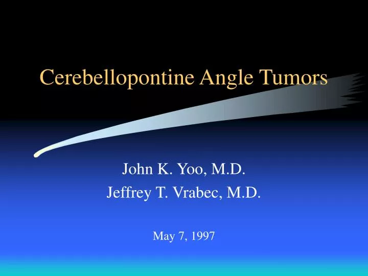
Cerebellopontine Angle Tumors. John K. Yoo, M.D. Jeffrey T. Vrabec, M.D. May 7, 1997. Anatomy. Borders of cerebellopontine angle Internal auditory canal Compartments of CN VII and VIII. History. Unilateral sensorineural hearing loss Sudden sensorineural hearing loss Unilateral tinnitus

E N D
Cerebellopontine Angle Tumors John K. Yoo, M.D. Jeffrey T. Vrabec, M.D. May 7, 1997
Anatomy • Borders of cerebellopontine angle • Internal auditory canal • Compartments of CN VII and VIII
History • Unilateral sensorineural hearing loss • Sudden sensorineural hearing loss • Unilateral tinnitus • Vestibular symptoms • Facial hypesthesia and weakness • Diplopia • Hoarseness, dysphagia, aspiration
Physical Examination • Thorough cranial nerve exam • Extra-ocular movements • Funduscopic exam • Facial motor and sensory function • Pneumatic otoscopy/Weber/Rinne • Hitselberger’s sign • Gag/TVC/SCM and trapezius
Diagnostic Tests • Pure tone and speech discrimination audiometry • Rollover • Impedance audiometry • acoustic reflex • tone decay • Auditory brainstem evoked response (ABR) • Vestibular testing (ENG)
Radiographic Studies • CT • MRI
Acoustic Neuroma • Benign slow growing tumors from Schwann cells surrounding CN VIII • 10% of the intracranial tumors and >90% of the CPA tumors • Incidence 0.1 to 2.5 per 100,000 • Associated with neurofibromatosis • Rate of growth 0.2 to 4.0 mm per year
Acoustic NeuromaRadiographic Image • Centered on IAC, spherical, enlarge the medial IAC, acute bone-tumor angle • CT: isodense and enhances with contrast • Inhomogeneous due to cystic degeneration or intratumoral hemorraging • MRI: isointense or hypointense on T1 and T2, but becomes markedly enhanced on T1-gadolinium
Acoustic NeuromaRationale of Management • Observation • Surgery for small intracanalicular tumors • Surgery for medium-sized tumors (1-3 cm) • Surgery for only-hearing ear • Surgery for bilateral acoustic neuromas (Neurofibromatosis-type II)
Meningiomas • 15% of intracranial tumors and 3% of CPA tumors • Arise from cells lining the arachnoid villa • Benign and do not metastasize, but locally aggressive because they invade bone • Signs and symptoms referable to site of involvement
MeningiomaRadiographic Image • Eccentric to IAC hyperostosis at medial IAC • Hemispherical and sessile with obtuse bone-tumor angle • CT: hypodense with calcification with marked enhancement; homogeneous • MRI: isointense/hypointense on T1, but only moderate enhancement on T1-gad
Meningiomas • Several histologic subtypes • syncytial • transitional • fibrous • angioblastic • sarcomatous • Surgical excision with removal of underlying bone
Hemangioma • Hamartomatous vascular malformations • Arise from geniculate ganglion or at the IAC • Closely associated with the facial nerve • MRI: hyperintense on T2 • CT: intratumoral bone spicules and “honeycomb” pattern of surrounding bone • Treatment is surgical excision
Other CPA lesions • Facial nerve schwannoma • Cholesteatoma (epidermoid) • Lipoma • Arachnoid cyst
Translabyrinthine • Advantages • No retraction of cerebellum • Allows good identification of CN VII • Allows good exposure of IAC • Less risk of CSF leak • Disadvantages • Hearing is sacrificed • Technique
Middle Fossa Approach • Advantages • Excellent for intracanalicular tumors, especially at the lateral end of the IAC • Hearing preservation is possible • Extradural with low risk of CSF leak • Disadvantages • Lack of access to CPA and posterior fossa • Need to retract temporal lobe • Technique
Retrosigmoid/SuboccipitalApproach • Advantages • Hearing preservation is possible • Access to CPA • Disadvantages • Limited access to lateral IAC/Fundus • Difficult to repairing or grafting CN VII • Increased risk of air embolism/CSF leak/ post-op headache • Cerebellar retraction is necessary • Technique