Download
1 / 85
880 likes | 1.02k Vues
Renal Disease. Dr. George Mellotte. The Kidneys Two bean-shaped organs, each the size of a fist. Weighing ~0.5% of total body weight 20% of Cardiac output goes to Kidney On Examination Move with Respiration Ballotable Can get above them Overlying resonance. Adrenal Glands. Rib Cage.

E N D
Renal Disease Dr. George Mellotte
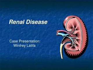
The Kidneys Two bean-shaped organs, each the size of a fist. Weighing ~0.5% of total body weight 20% of Cardiac output goes to Kidney On Examination Move with Respiration Ballotable Can get above them Overlying resonance Adrenal Glands Rib Cage Kidneys Rib Cage Bladder
Function of the Kidney • Primary balancing organ needed to keep blood in a stable state • Remove waste products and toxins • Urea & Creatinine: • Drugs, toxic substances • Maintain fluid and electrolyte balance • Total body water and fluid distribution • 50-70% of body weight is water • Sodium Potassium • Maintain normal mineral balance • Calcium Phosphate Magnesium • Regulate acid/base balance
Function of the Kidney • Endocrine Role of the Kidney • Regulates blood pressure: Renin-angiotension System and aldosterone • Adjusts final concentration of urine Antidiuretichormone ADH • Stimulates the production of red blood cells Erythropoietin • Activates Vitamin D (Calcitriol, 1,25(OH)2D3) • Response to Parathyroid hormone
Target Organ Damage • Heart • Left ventricular hypertrophy • Angina or prior myocardial infarction • Prior coronary revascularization • Heart failure • Brain • Stroke or transient ischemic attack • Chronic kidney disease • Peripheral arterial disease • Retinopathy
Estimating Renal Function • Serum creatinine is widely used BUT • Serum creatinine is based on muscle mass • ‘Normal’ values are lower for elderly, female or physically inactive patients • During early nephron loss, adaptive changes compensate to minimise rise in creatinine What is normal? • Newer evidence suggests lower thresholds should be used, especially for women Creatinine : Men 50 - 115 umol/l Women 40 - 95 umol/l
Defining Renal Failure Cr Clearance = (140 - age) × weight in Kg × SF serum Cr SF = 1.2 males / 1.05 females Normal GFR = 90-120mls/min GradeGFRSr CreatinineMild 60 - 90ml/min 100 - 150umol/l Moderate 30 - 60ml/min 150 - 250µmol/lSevere 15 - 30ml/min 250 - 500µmol/l Endstage < 15ml/min > 500µmol/LCockcroft & Gault Formula
Classification of Chronic kidney disease (CKD) Description GFR (mL/min/1.73 m2) Stage 1 Kidney damage with normal / GFR 90 2 Kidney damage mildGFR 60–89 3 Kidney damage Moderate GFR 30–59 4 Kidney damage Severe GFR 15–29 5 End Stage Kidney failure < 15 (or dialysis) eGFR can be thought of as equivalent to % kidney Function www.kidney.org/professionals/kdoqi GFR, glomerular filtration rate
Key Concepts • The importance of early identification • Kidney Disease • Cardiovascular Disease • Focus on quality of care before starting Dialysis • Slowing the progression of Kidney Disease • Slowing the progression of Co - Morbid Disease • Interplay of pathophysiology • Progressive Kidney Disease • Progressive Cardiovascular Disease
JNC 7 Report on Hypertension • For persons over age 50, SBP is a more important than DBP as CVD risk factor. • Starting at 115/75 mmHg, CVD risk doubles with each increment of 20/10 mmHg throughout the BP range. • The BP relationship to risk of CVD is continuous, consistent, and independent of other risk factors
New Features and Key Messages • Thiazide-type diuretics should be initial drug therapy for most, either alone or combined with other drug classes. • Certain high-risk conditions are compelling indications for other drug classes. • Most patients will require two or more antihypertensive drugs to achieve goal BP. • If BP is >20/10 mmHg above goal, initiate therapy with two agents, one usually should be a thiazide-type diuretic.
All Patients with nephropathy – particularly diabetic patients Target BP = 125/75 mmHg Re proteinuria - a lower BP is permissible/desirable in order to maximise ACEI & ARB – Target Proteinuria <0.3g/day
Benefits of Lowering BP Average Percent Reduction Stroke incidence 35–40% Myocardial infarction 20–25% Heart failure 50% In stage 1 HTN and additional CVD risk factors, achieving a sustained 12 mmHg reduction in SBP over 10 years will prevent 1 death for every 11 patients treated.
Ambulatory BP Monitoring • ABPM is warranted for evaluation of “white-coat” HTN in the absence of target organ injury. • Ambulatory BP values are usually lower than clinic readings. • Awake, individuals with hypertension have an average BP of >135/85 mmHg and during sleep >120/75 mmHg. • BP drops by 10 to 20% during the night; if not, signals possible increased risk for cardiovascular events.
CVD Risk Factors • Obesity (BMI >30 kg/m2) Physical inactivity • Cigarette smoking • Hypertension Hyperlipidaemia • Diabetes mellitus • Microalbuminuria Proteinuria • eGFR <60 ml/min Mild Renal failure Creatinine>125 • Age (older than 55 for men, 65 for women) • Family history of premature CVD (men under age 55 or women under age 65) *Components of the metabolic syndrome.
Identifiable Causes of Hypertension • Chronic kidney disease • Renovascular disease • Primary aldosteronism • Sleep apnea • Drug-induced or related causes • Chronic steroid therapy and Cushing’s syndrome • Pheochromocytoma • Coarctation of the aorta • Thyroid or parathyroid disease
Laboratory Tests • Routine Tests • ECG • Urinalysis • Blood glucose, and haemoglobin • Renal profile (potassium, creatinine, calcium) • Lipid profile, • Hypokalaemia without diuretics – secondary cause? • Optional tests • Microalbuminuria or albumin/creatinine ratio • Renal Ultrasound if renal impairment • More extensive testing for identifiable causes is not generally indicated unless BP control is not achieved
Minority Populations • In general, treatment similar for all demographic groups. • Socioeconomic factors and lifestyle important barriers to BP control. • Prevalence, severity of HTN increased in African Americans. • African Americans demonstrate somewhat reduced BP responses to monotherapy with BBs, ACEIs, or ARBs compared to diuretics or CCBs. • These differences usually eliminated by adding adequate doses of a diuretic.
Risk Factors for Vascular Disease in CKD 1 • Traditional Risk Factors • Hypertension • Left Ventricular Hypertrophy • Hyperlipidaemia • DOQI Guidelines – Treat similar to Diabetes/Post MI • Total Cholesterol < 4.0 mmol/l • LDL Cholesterol < 2.5 mmol/l • Will require statins – • Beware of the patient with Hypocholesterolaemia • Marker of nutritional deficiency & of increased mortality risk
Risk Factors for Vascular Disease in Renal Failure 2 • Renal Specific Risk Factors • Proteinuria • Anaemia • Hyperhomocysteinaemia • Hyperphosphataemia • Uremia per se?
Main Factors in Progression of Renal Failure • Genetic / Racial determinants • Age of onset of nephropathy • Sex • Underlying Disease • Co-Morbid Disease • Blood Pressure control • Degree of Urinary Protein Excretion
Progression in Renal Failure Destruction of Nephrons Destruction of Nephrons Remaining Nephrons Focal & Segmental Glomerulosclerosis Glomerular Hyperfiltration
Renal damage induces hypertension via • Plasma volume expansion, • Sodium retention, • Overactivity of both the sympathetic nervous system and the renin-angiotensin-aldosterone axis, • Accumulation of circulating endogenous vasoactive substances. “Early CRF typically results in a 10-20 mm Hg increase in diastolic blood pressure until, and unless, renal impairment is identified and treated” Lancet 2000; 356 147-52
Relationship between achieved BP control and declines in GFR in clinical trials of diabetic & non-diabetic renal disease
Blockade of the renin angiotension system is ‘RENOPROTECTIVE’ • Evidence based medicine suggests preferred initial therapy is either an ACE Inhibitor or Angiotension II Receptor Blocker 3 • Most trials note a 24%- 50% risk reduction of overt nephropathy, independent of BP reduction • Recent evidence suggests a synergy between ACE inhibitors and AII Blockers
Acute Renal Failure (ARF) A clinical condition – defined as an abrupt rise in Urea & Creatinine (with or without Oligiouria) • 3 - 6 % of all hospital admissions develop ARF • Incidence higher in complicated cases such as seen in Cardiac surgery • IncidenceARFS (Acute Renal Failure Scotland) Study • Rise in Creatinine requiring intervention 237.9 pmp/year • Incidence requiring dialysis 118.7 pmp/year • 60% treated in ICU • 33% had preexisting renal disease • Development of ARF increases the risk of death associated with a particular procedure - 5 fold
Pathophysiology of ARF • Not simply a renal hypoperfusion problem • Restoration of renal blood flow would correct urine flow • Altered renal blood flow post ischaemic insult • Changes in renal vasoconstriction/vasodilation • Pooling of blood in Renal medulla • Changes in inflammatory markers • Increased leukocyte adhesion • Increased pro-inflammatory markers (TNF, IL6) • Increased pro-coagulation activation • Tubular Alterations • Effects of cytoskeletal Breakdown – loss of polarity
Avoiding Renal Failure • Prevent Dehydration in High Risk Patients • Patients with S. Creatinine >125umol/l (?lower in females) • Patients with Diabetes, Peripheral Vascular Disease or Myeloma • Controlled trials recommend N. Saline @ 1ml/kg/hr 12 hrs pre & post procedures – • particularly important pre Major Surgery or where contrast will be administered • CRF patients have a fixed urinary concentrating deficit • Role for N-Acetyl Cysteine to prevent contrast Nephropathy • Adjust drug dosing where there is renal impairment • Renal tubules more sensitive to the effect of nephrotoxins in the presence of renal hypoxia
Guidelines for immediate management of patients with oliguria or anuria • Assess & correct any respiratory or circulatory impairment • Manage any life threatening consequences of renal dysfunction (hyperkalaemia, salt and water overload, extreme acidosis) • Exclude obstruction of the urinary tract - Get Ultrasound • Establish underlying cause(s) and institute prompt remedial action • Get a drug history and alter prescriptions appropriately • Get help from senior appropriately trained specialists
Initial Investigations • Blood Tests FBC, blood film, Coag screen, U/E, LFT's, Bone profile, ABG, Immunology if appropriate- ANA, DsDNA, C3,C4, CRP, SPEP, ANCA & Anti GBM ASOT titre • Urine Electrolytes & Osmolarity Urine Microscopy • X-ray Renal Ultrasound Isotope Perfusion scan
Diagnostic Imaging • Plain films (K.U.B.)Screening tool for renal stones. renal calcification • Ultrasoundsafe, high quality images can be obtained on most patientsGood screening tool – esp in ARFUsed to evaluate renal size, renal masses and obstruction • Intravenous Pyelography (I.V.P.)assesses the collecting system and urinary tract Increasingly being replaced by CT-IVP & MRI Not for patients at risk of contrast nephropathy - Renal failure, Multiple Myeloma, Diabetics Volume depleted patients • Nuclear Medicine – RenogramUsed to asses renal function - DTPA or MAG3 • MRI • Increasingly being used in general nephrology • MR Angiography • MR Urography
Pre-Renal Vs Established Renal failure Pre-renal RF Inadequate renal perfusion The kidneys concentrating power is normal and the urine produced is highly concentrated Established RF Failure of tubular function The kidney’s concentrating power is severely damaged and the urine produced is dilute Early restoration of effective circulation will avert ATN (Acute Tubular Necrosis)
Renal Hypoperfusion V Established ARF MeasurementPre Renal ATN Urinary Na (mmol/l) <20 >40 Fractional excretion <1 >4of Na (%) Only appropriate if diuretics not given
Restore Renal Perfusion Correct Hypovolaemia Target CVP = 10 cm Correct HypotensionTarget MAP > 75mmHg Use fluids in first instance Start Inotropic support if response insufficient No Diuresis Frusemide 100 - 250mg IVPt must be euvolaemicê Diuresis Measure hourly urine output & replace losses Management of Pre Renal Failure Lack of response indicates that ATN has developed
Diuretic therapy Ineffective once ATN is established Inappropriate in inadequately treated prerenal Will require a higher dose to achieve effect If overloaded Diuresis should not be at the expense of hypotension Aim negative balance 0.5 – 1L day unless clinically indicated Replacement for even balance is Positive 0.5L for insensible losses This is true irrespective of urine output! Guidelines for fluid management in ARF
Damage Decrease in renal perfusion Impaired intrarenal haemodynamics Tubular toxicity Allergic interstitial nephritis Class of drug Diuretics, ACE inhibitors, B-Blockers, vasodilators NSAID’s, radiocontrast Aminoglycosides, amphotericin, cisplatin lactams,(penicillins) NSAID’s Drugs that induce renal damage
ECG changes of Hyperkalaemia • Peaked T waves, Flattened P wave • Prolonged PR interval è sinus arrest • Wide QRS complexes & deep S waves èSine Wave èV. Fib è asystole
Management of Hyperkalaemia Hyperkalaemia is a medical emergency and must be corrected immediately. • V. Fib likely if K+ > 7.0 mmol/l (in ARF) Rx 1. 10-20 mls of 10% Calcium Gluconate 2. 50 mls of 50% Dextrose with 12 IU Insulin over 30 mins followed by infusion @ 10ml/hour 3. 50 - 100 mls of 8.4% NaHCO3 if acidotic
Indications for renal replacement therapy • Uncontrollable hyperkalaemia (K+ > 6.5) • Salt and water overload unresponsive to diuretics • Anuric and/or need to administer fluid/feed • Severe uraemia (Creat >500umol/l ARF) • Acidaemia Consider if pH <7.2 Multiple indications may trigger earlier intervention
Chronic Renal Failure • Defined as permanent loss of renal function • Prevalence underestimated • In USA - while only 0.1% of population require dialysis 5-10 % have renal dsease • Most patients have no symptoms until CRF is advanced • Advanced CRF often termed End Stage Renal Disease (ESRD) • Definedas a GFR <15mls/min • Typical symptoms are nausea, anorexia, fatigue, itch and bruising • Typical signs are hypertension, ankle swelling, breathlessness and anaemia
Chronic Renal Failure • Laboratory features • Urea & Creatinine • Hyperkalaemia • Hypocalcaemia • Hyperphosphataemia • Metabolic Acidosis • Normochromic Anaemia • Radiology - Ultrasound • Small kidneys - often scarred Clinical features Symptoms • Nausea Vomiting • Fatigue Obtunded • Pruritus Bruising Signs • Hypertension • Oedema CCF • Anaemia • Pericarditis - late • Neuropathy - late
Progression of Renal Disease • Progression in CRF due in part to secondary factors unrelated to the activity of the initial disease • focal segmental glomerulosclerosisProteinuria • Good evidence to support strategies to minimise amount of proteinuria • Chiefly - ACE inhibitors - BP control - Diabetic control
Haemodialysis Intermittent Complex Hospital Based Problems Poorly tolerated in cardiac disease Vascular access Most suitable Active patients Patients with limited ability to self care Nephrotic Syndrome Transplantation Desirable Scarce resource Not a cure Problems Graft Failure Infection risk Cancer Risk Only medically fit patients are Transplanted Treatment Choice C.A.P.D. /A.P.D. Simple Independent Patient dependent Problems Peritonitis risk Catheter Malfunction Protein losses Most Suitable Diabetics Elderly Patients living away from aHD unit
When to start Dialysis • Accepted reasons • Patient has symptoms of uraemia • Kidney function approx 15% of predicted (GFR < 10-15mls/min) • Can wait until lower if patient remains well • Patient develops high serum potassium levels • The need to start Dialysis can often be delayed using Erythropoietin (EPO) to minimise symptoms • Controversial - does “early start” benefit patient? • ?less malnutrition ?less cardiac damage