Download

1 / 35
360 likes | 926 Vues
Benign Paroxysmal Positional Vertigo. Amy Stinson MS IV Kansas City University of Medicine. BPPV. General Considerations History Anatomy Pathogenesis Clinical Evaluation Treatment Prognosis. BPPV. Most common cause of peripheral vertigo

E N D
Benign Paroxysmal Positional Vertigo Amy Stinson MS IV Kansas City University of Medicine
BPPV • General Considerations • History • Anatomy • Pathogenesis • Clinical Evaluation • Treatment • Prognosis
BPPV • Most common cause of peripheral vertigo • Most common identifiable cause – Head trauma, 2nd – vestibular neuronitis • Predisposing factors: infection, surgery, prolonged bed rest, Meniere’s disease • Usually idiopathic 50 – 70% • Incidence 64:100,000 every year • 20-30% of diagnosed vertigo
History • 1921 – Barany • 1952 – Dix and Hallpike • 1969 – Schuknecht • Proposed posterior canal crista was source of dysfunction • Loose otoconia from utricle deposited on cupula • Cupulolithiasis • Concluded that ampullofugal (excitatory) deflection of posterior canal cupula accounts for nystagmus
History • 1979 - Hall, Ruby & McClure • BPPV results from deflection of posterior canal cupula because of the motion of debris within the posterior canal • Canalithiasis • This accounted for fatigability of nystagmus • 1985 – McClure – horizontal canal BPPV • 1994 – Brandt – anterior canal BPPV
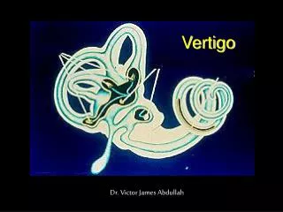
Anatomy • Vestibular portion of CN 8 arises in Scarpa’s ganglion in internal auditory meatus • Peripheral processes of bipolar ganglion cells terminate in hair cells of sensory epithelium of the labyrinth • Hair cells sit on the surface of cristae • Cristae ampullaris – SCC • Maculae acousticae – utricle and saccule • Hair cells are covered by: • Cupula - SCC • Otolithic membrane - maculae
Brodel M: Three unpublished drawings of the anatomy of the human ear, Philadelphia, WB Saunders, 1946
Maculae Acousticae of Utricle and Saccule
Semicircular canals Ampullae senses head turning – angular acceleration Endolymph w/in canal causes cupula to move deflection of hair cells sensation of rotation Utricle and Saccule Maculae senses gravity and head tilt – linear acceleration Hairs are displaced in response to gravity on otoliths sensation of tilt Anatomy
Anatomy Barber HO, Stockwell CW: Manual of electronystagmography, St Louis, Mosby, 1976
Pathogenesis • Canalolithiasis • Most widely accepted hypothesis of BPPV • Otoconia become displaced from utricular macula. Because the particles are heavier than surrounding endolymph, they tend to collect in the long arm of the posterior semicircular canal. • Once the particles clump into a sufficient mass, changes in head position cause gravitation of the particles hydrodynamic drag on the endolymph displacing the cupula
Pathogenesis • 5 Typical Features of PC –BPPV • 1. The canalithiasis mechanism explains the latency of nystagmus as a result of the time needed for motion of the material within the posterior canal to be initiated by gravity • 2. The nystagmus duration is correlated with the length of time required for the dense material to reach the lowest part of the canal • 3. The upbeating (vertical) and torsional components of nystagmus are consistent with eye movements evoked by stimulation of the posterior canal nerve
Pathogenesis • 4. The reversal of nystagmus when the patient returns to sitting upright position is due to retrograde movement of particles in PC lumen back towards the ampulla • 5. The fatigability of nystagmus evoked by repeat Dix-Hallpike positional testing is explained by dispersion of particles within the canal
Pathogenesis • Horizontal(Lateral) Canal – BPPV • 2 - 15% BPPV pts • Idiopathic, minor head trauma, complication of Tx of PC-BPPV • Turning the head while supine evokes severe vertigo • Cupulolithiasis plays a greater role • Resulting nystagmus is horizontal • Geotropic – toward undermost ear • Apogeotropic – beats away from undermost ear (rarer)
Pathogenesis • Anterior Canal – BPPV • Similar provoking factors as LC and HC – BPPV • Nystagmus is downbeat and torsional • Latency, duration & fatigability are similar
Case • 69 yo female c/o several months of episodic dizziness described as spinning and imbalance associated with severe nausea • Last episode occurred when she got out of bed and felt dizzy within seconds • She has awakened from sleep with a swimming sensation • She has had spinning sensations lasting less than a minute when reaching into an upper cupboard • Pt admits to being a “fender bender” a few months ago while snowbirding down in Florida
Case • Exam is normal except for paroxysmal positional upbeating and counterclockwise torsional nystagmus with Dix-Hallpike positioning to the right side • Canalith repositioning is performed with resolution of her nystagmus upon repeat positioning
Clinical Evaluation • 50 y/o Female • Recurrent episodes of vertigo lasting less than one minute (usually a few seconds) • Associated with change in head position • Nausea and vomiting • Symptoms may fatigue as day progresses • Episodes can continue for weeks to months
Clinical Evaluation • Normal neurologic exam • Normal hearing test and tympanogram • No spontaneous nystagmus • Dix-Hallpike test • 1-2 sec latency of onset of vertigo and nystagmus • Nystagmus is classically torsional (rotatory) with vertical component (counterclockwise for right ear and clockwise for left ear) • Nystagmus is fatigable with repeated tests
Clinical evaluation • Roll test • Log roll or barbeque test • Supine head turning elicits horizontal (lateral) canal BPPV • Anterior canal BPPV most commonly spontaneously resolves
Treatment • Repositioning maneuvers • Epley – effective in over 90% of cases • Most effective for PC-BPPV • Sermont – more difficult to perform • No advantage over Epley • After maneuvers, pts should avoid bending over and should sleep with their head elevated at least 45° for the next 48 hrs
Treatment • Surgical • Singular neurectomy – • For Highly intractable BPPV • The post. ampullar br. of vestibular nerve is transected just before it enters the amuplla • Complete resolution in 80 – 97% of pts • Sensorineural hearing loss 4 – 6%
Treatment • Surgical • Posterior Semicircular Canal Occlusion • Obstruction of canal lumen preventing the flow of endolymph • This fixes the cupula and renders it unresponsive to angular acceleration • Post-op imbalance and disequilibrium and transient sensorineural loss that usually resolves within a few weeks
Prognosis • Natural history of BPPV includes acute onset and remission over a few months • 90 – 95% of pts will respond to one repositioning maneuver • Pts can have unpredictable recurrences that often respond to a repositioning maneuver • With intractable disease posterior canal occlusion is safe and reasonable option
References • Cummings: Otolaryngology: Head & Neck Surgery, 4th Ed. • UpToDate: Positional vertigo and nystagmus • Fife, TD. Recurrent positional vertigo. Continuum: Lifelong learning in neurology. Aug 2006. 12:92-115. • Quinn, FB. Ryan, MW. Medical management of vestibular disorders and vestibular rehabilitation. Grand rounds, UTMB Dept. of Otolaryngology. 2004. • Adams and Victor’s Neurology. Deafness, Dizziness, and Disorders of equilibrium. Chap 15. 2006. • Lange Neurology. Disorders of Equilibrium. Peripheral vestibular disorders. Chap 3. 2006. • Lange. Current Diagnosis and treatment of Otolaryngology – Head and neck surgery. Vestibular system. Chap 43. 2004. • Shaia, WT et al. Success of Posterior Semicircular Canal Occlusion and Application of the dizziness Handicap Inventory. Otolaryngology – Head and neck surgery. 2006. 134:424-430. • White, JA. Oas, JG. Diagnosis and Management of Lateral Semicircular Canal Conversions during Particle Repositioning Therapy. Laryngoscope. 2005. 115:1895-1897. • Virre, E. Purcell, I. The Dix-Hallpike Test and the Canalith Repositioning Maneuver. Laryngoscope. 2005. 115:184-187. • Woodworth, BA. Et al. The Canalith Repositioning Procedure for Benign Positional Vertigo: a Meta-Analysis. Laryngoscope. 2004. 114:1143-1146.
References • Kos, MI. Et al. Transcanal approach to the Singular Nerve. Otology and Neurotology. 2006. 27:542-546. • Parnes, LS. Agrawal, SK. Diagnosis and management of benign paroxysmal positional vertigo. CMAJ. 2003. 169:681-693. • Walsh, RM. Bath, AP. Cullen, JR. Long-tern results of posterior semicircular canal occlusion for intractable benign paroxysmal positional vertigo. Clinical Otolaryngology & Allied Sciences. 1999. 24:316-323. • Sekine, K. Imai, T. et al. Natural History of benign paroxysmal positional vertigo and efficacy of Epley and Lempert maneuvers. Otolaryngology – Head & Neck Surgery. 2006. 135:529-533. • White, JA. Coale, KD. Diagnosis and management of lateral semicircular canal benign paroxysmal positional vertigo. Otolaryngology – Head & Neck Surgery. 2005. 133:278-284. • Korres, SG. Diagnostic. Pathophysiology, and therapeutic aspects of benign paroxysmal positional vertigo. Otolaryngology – Head & Neck Surgery. 2004. 131:438-44.