Download
1 / 42
470 likes | 876 Vues
IMAGE CREATION. ATOMSINTERACTION WITH

E N D
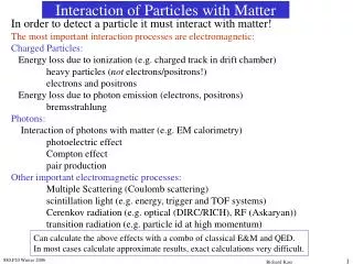
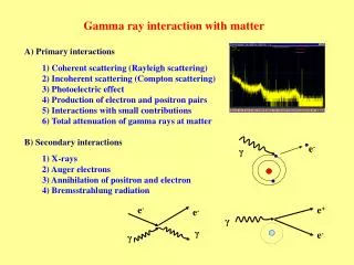
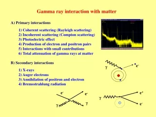
1. X-Ray Interaction with Matter & Human Biology
4. Patient Interactions **Photoelectric**
Classic Coherent Scatter
**Compton Scattering**
Pair Production
Photodisintegration
9. Coherent Scattering The wavelength is equal to the incident x-ray or equal energy.
The only difference is the direction of travel
Energy in = Energy out - Only changes is direction
11. Compton Effect or Compton Scattering Occurs throughout the diagnostic imaging range
The incident x-ray interacts with the outer electron shell on an atom of matter, removing it.
It not only causes ionization but scatters the incident x-ray causing a reductions in energy and the change of direction.
12. Compton scatter A fairly high energy (high kVp) x-ray photon ejects an outer shell electron.
Though the x-ray photon is deflected with somewhat reduced energy (modified scatter), it retains most of its original energy and exits the body as an energetic scattered photon.
A Compton e- is also released
Since the scattered photon exits the body, it does not pose a radiation hazard to the patient.
It can, however, contribute to film fog and pose a radiation hazard to personnel (as in fluoroscopic procedures).
15. Compton scatter Both the scattered x-ray and the Compton electron have enough energy to cause more ionization before loosing all their energy
In the end the scattered photon is absorbed photoelectrically
16. Compton Effect The Compton electron looses all of its kinetic energy by ionization and excitation and drops into a vacancy in an electron shell previously created by some other ionizing event
The probability of Compton effect increases as photon energy increases, however the atomic number does not affect the chances of the Compton effect
17. Compton Scatter Compton is just as likely to occur with soft tissue as bone. Compton can occur with any given photon in any tissue
Compton is very important in Radiography, but not in a good way.
Scattered photons provides no useful diagnostic information
18. Compton Effect Scattered radiation produces a uniform optical density on the radiograph that reduces image contrast
Scattered radiation from Compton contributes to the majority of technologists exposure, especially during fluoroscopy
STAY AWAY FROM YOUR PATIENT !
19. Scatter from the Patientduring Fluoroscopy
20. ISOEXPOSURE CURVES
21. Photoelectric Effect or Absorption Inner-shell ionization
The photon is not scattered it is totally absorbed
The e- removed from the atom of matter is called a photoelectron, with an energy level equal to the difference between the incident photon and the e- binding energy.
22. Binding Energy is very important Table 10-2
28. Photodisintegration
29. PHOTOELECTRIC
ABSORBTION
IS WHAT GIVES US
THE CONTRAST
ON THE FILM
31. Compton scatter Contributes to no useful information
Is independent of the atomic number of tissue. The probability of Compton is the same for bone atoms and for soft tissue atoms
The probability for Compton is more dependent on kVp or x-ray energy
32. Compton Scatter Results in image fog by optical densities not representing diagnostic information
Photon are Photons
IR is does not know
the difference
33. Photoelectric Absorption Provides information to the IR because photons do not reach the IR
This represents anatomic structures with high x-ray absorption characteristics; radiopaque structures; tissue with high atomic number; or tissue with high mass density
34. Attenuation � The total reduction in the # of photons remaining in an x-ray beam after penetration through tissue Absorption = x-ray disappears (Photoelectric, Pair production & Photodisintegration)
Scattering = partially absorbed, x-ray emerges from the interaction traveling in a different direction (sometimes with less energy)
Absorption + Scattering = Attenuation
35. 3 Types of x-rays are important for IMAGE FORMATION DIFFERENTIAL ABSORPTION = the difference between those x-rays absorbed and those transmitted to the IR
Compton scatter (no useful information)
Photoelectric absorption (produces the light areas on the image)
Transmitted x-rays (produces the grey/dark areas on the image)
38. Differential Absorption The difference in x-ray interactions
Fundamental for image formation
Occurs because of Compton Scattering, Photoelectric absorption, and X-ray transmission
39. Differential Absorption
40. Compton vs. Photoelectric Below 60 kVp Photoelectric absorption is predominant above 60 kVp Compton scatter begins to increase.
Dependent on the tissue attenuation properties
Table 10-13
41. Differential absorption factors High atomic number = larger atoms
Mass Density = how tightly the atoms of tissue are packed
Z # for air and soft tissue are about the same the OD changes are due to mass density difference
Table 12�3 & 12-5
42. Radiation Protection Producing high-quality radiographs require careful technique selection, reducing kVp improves differential absorption and image contrast
However, patient dose is increased because more photons are absorbed by the body