Download
1 / 60
740 likes | 1.41k Vues
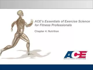
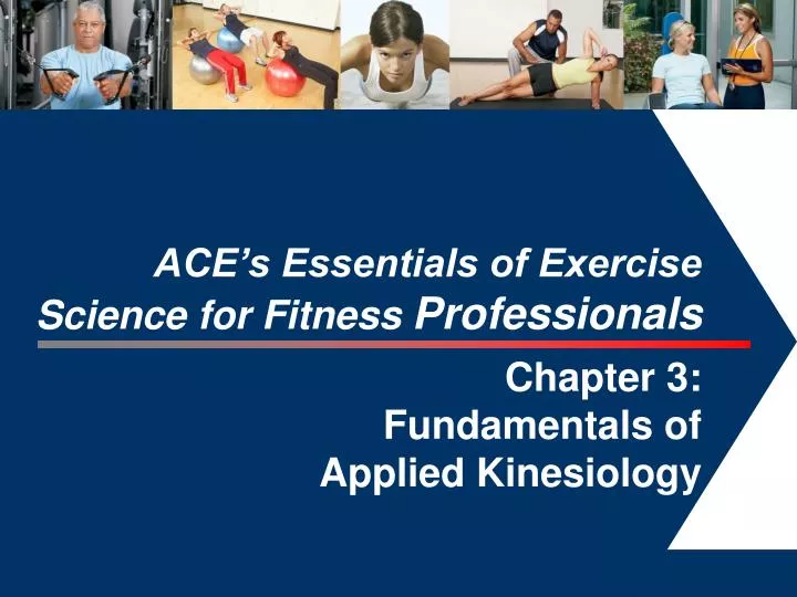
ACE’s Essentials of Exercise Science for Fitness Professionals Chapter 3: Fundamentals of Applied Kinesiology. 1. Learning Objectives.

E N D
ACE’s Essentials of Exercise Science for Fitness Professionals Chapter 3: Fundamentals of Applied Kinesiology 1
Learning Objectives • This session, which is based on Chapter 3 of ACE’s Essentials of Exercise Science for Fitness Professionals, explains the functional kinesiology of the upper extremity, lower extremity, and spine and pelvis. • After completing this session, you will have a better understanding of: • Biomechanical principles applied to human movement • The kinesiology and muscle function of the lower extremity • The kinesiology and muscle function of the upper extremity • Exercises to strengthen and stretch key muscle groups • Obesity and age-related considerations
Introduction • Kinesiology involves the study of human movement from biological and physical science perspectives. Understanding the principles and concepts of human movement provides the framework to analyze movement and design safe and effective programs for clients and class participants. • This understanding will also provide a foundation for identifying and addressing: • Proper body mechanics • Neutral postural alignment • Muscular balance
Newton’s Laws of Motion • Newton’s laws of motion describe the interrelationships among force, mass, and human movement and are applied at either individual joints or the body as a whole. • Law of inertia: A body at rest will stay at rest and a body in motion will stay in motion (with the same direction and velocity) unless acted upon by an external force. • Law of acceleration: Force (F) acting on a body in a given direction is equal to the body’s mass (m) multiplied by its acceleration (a). • Law of reaction: Every applied force is accompanied by an equal and opposite reaction.
Types of Motion • Motion is a change in an object’s position in relation to another object. • There are four basic types of motion: 1. Rotary: An object is fixed and turns around a fixed point (angular motion) Rotary motion. Each point in the forearm/hand segment follows the same angle, at a constant distance from the axis of rotation (A), and at the same time.
Types of Motion (cont.) Translatory motion. Each point on the forearm/hand segment moves in a parallel path through the same distance at the same time. 2. Translatory: An object is not fixed and moves in a straight line (linear) 3. Curvilinear: A small gliding motion within a joint (linear or translatory) combined with rotary motion of a segment 4. General plane motion: Motions at various joints are simultaneously linear and rotary.
Forces • Force is a push or pull exerted by one object on another. • External force • Muscular contractions • Human movement is often described in terms of motive and resistive forces. • Motive force causes an increase in speed or a change in direction. • Resistive force resists the motion of another external force.
Muscular Actions • Concentric contraction • Muscle acts as the motive force and shortens as it create tension. • Motion is created by the muscle contraction. • Eccentric contraction • Muscle acts as the resistive force and lengthens as it creates tension. • External force exceeds the contractive force generated by the muscle. • Motion is controlled (slowed) by the muscle contraction. • Isometric contraction • Muscle tension is created, but there is no apparent change in length. • Resistance can come from opposing muscle groups, gravity, an immovable object, or weight training. • Motion is prevented by the muscle contraction (equal opposing forces)
Levers • A lever is a rigid bar with a fixed point (fulcrum) around which it rotates when an external force is applied. • Rotary motion occurs in one of the three planes of motion, where the axis of rotation (which intersects the center of the joint) is perpendicular to the plane. • Sagittal plane: mediolateral axis • Frontal plane: anteroposterior axis • Transverse plane: longitudinal axis
Torque • For rotation to occur, the motive force must contact the lever at some distance from the axis of rotation. • Torque is the turning effect that occurs when the force acts on the lever arm. • The pull of the biceps brachii on the radius creates a third-class level with axis of rotation at the elbow joint.
Lever Classes • There are three lever classes. • The body operates primarily as a series of third-class levers, with only a few first- and second-class levers. • Force (F) acts between the axis (X) and the resistance (R)
Third-class Levers in the Body • In a third-class lever, the motive force has a short lever arm and the resistance has a long lever arm. • Motive force muscles are at a mechanical disadvantage. • Muscles typically attach near the joint, creating a short lever arm and, as a result, it requires relative high forces to lift even small weights. • Application to training: • Assuming a client is lifting the same amount of weight, he or she can create more resistance by moving the weight farther from the working joint, or less resistance by moving it closer to the working joint.
Muscle Fiber Arrangements • In addition to neurological training and recruitment, muscle fiber type, number, size, and arrangement influence a muscle’s ability to create force. • Muscle fiber arrangements include: • Penniform (unipennate, bipennate, multipennate) • Longitudinal
Human Motion Terminology • Terms for muscle function and the roles that muscles play during movement: • An agonist (or prime mover) is a muscle that causes a desired motion, while antagonists are muscles that have the potential to oppose the action of the agonist. • Synergist muscles assist the agonist in causing a desired action. • Co-contraction describes when the agonist and antagonist contract together and a joint must be stabilized.
Kinetic Chain Movement • Optimal performance of movement requires that the body’s muscles work together to produce force while simultaneously stabilizing the joints. • Closed-chain vs. open-chain movement • Closed-chain movement: The end of the chain farthest from the body is fixed. This type of movement emphasizes stabilization through joint compression and muscle co-contraction. • Open-chain movement: The end of the chain farthest from the body is free. This type of movement involves more shearing forces at joints. • A joint’s mobility (range of uninhibited movement) should not be achieved by compromising joint stability.
Balance and Alignment of the Body • Center of gravity (COG) • The point at which a body’s mass is considered to concentrate and where it is balanced on either side in all planes (frontal, sagittal, and transverse) • Also the point where gravity is enacting its constant downward pull
Line of Gravity and Base of Support • Gravity acts on the body in a straight line through its COG toward the center of the earth—called the line of gravity. • To maintain balance without moving, the line of gravity must fall within the base of support (BOS). • The BOS is the area beneath the body that is encompassed when one continuous line connects all points of the body that are in contact with the ground. • Balanced, neutral alignment requires that the body parts are equally distributed about the line of gravity within the BOS.
Gravity and Muscular Actions The primary muscles must contract concentrically to lift an object or create human movement that is in a direction opposite the pull of gravity. The primary muscles must contract eccentrically to lower an object or control human movement that is in the same direction as the pull of gravity. If gravity is eliminated [e.g., in movements being performed perpendicular to the pull of gravity (parallel to the floor)], each muscle group acts concentrically to produce the movement.
Anterior Hip Muscles: Hip Flexors • Active range of motion for hip flexion • Prime movers: iliopsoas, rectus femoris, sartorius, pectineus, and tensor fasciae latae • Act synergistically to cause hip flexion (e.g., “up” phase of a knee lift) • Act eccentrically to control hip extension (e.g., “down” phase of a knee lift)
Hip Flexors: Considerations • Muscle origins and insertions impact muscular function. • Iliopsoas • The psoas major and minor originate in the low back and insert to the proximal femur, leading to poor mechanical leverage when used to raise and lower a straight leg. The abdominals are not strong enough to balance the large force and keep the spine in neutral alignment. Sample strengthening exercise: Sample stretching exercise:
Hip Flexors: Considerations • Rectus femoris • Works at both the knee and hip, concentrically contracting to perform hip flexion and knee extension. • Sample strengthening exercise: standing straight-leg raise • Sample stretching exercise: iliopsoas lunge, bending the back knee • The sartorius is the longest muscle in the body. It is also involved in hip abduction, adduction, and external rotation, and knee flexion and internal rotation. • Tensor fascia latae (TFL) • IT band • Explosive hip flexion results in highly developed TFL
Posterior Hip Muscles: Hip Extensors • Active range of motion for hip extension • Prime movers: hamstrings (biceps femoris, semitendinosus, semimembranosus) and gluteus maximus • Activated concentrically to extend the hip joint (e.g., prone leg lift) • Activated eccentrically to control hip flexion (e.g., downward phase of squat)
Hip Extensors: Considerations • The hamstrings work as prime movers during normal walking and low-intensity activity. • The gluteus maximus is a prime mover during higher-intensity activities such as stair climbing, sprinting, and stationary cycling. • Higher-intensity activities typically require greater hip ranges of motion and more powerful extension. • Guideline for activities that involve the gluteus maximus: Choose hip extension exercises that require at least 90 degree of hip flexion at the start of the hip extension movement.
Lateral Hip Muscles: Hip Abductors • Active range of motion for hip abduction • Prime movers: gluteus medius, gluteus minimus, and superior fibers of gluteus maximus • Assisted by the TFL • Act concentrically to abduct the hip • Two-thirds of the gluteus maximus muscle fibers cross inferior to the joint axis, making them involved in hip abduction and adduction
Hip Abductors: Considerations Side-lying leg lifts (upper leg): abductors work concentrically in the upward phase and eccentrically in the downward phase Supine hip abduction/adduction with the hips extended: abduction of the hip joints occurs as the legs move further apart, while the adductors control the movement of the legs together Concentric (legs apart) action of the hip abductors with elastic resistance When the hip is flexed more than 40 degrees, the six external rotators become the prime movers of hip abduction. Strengthening exercises:
Lateral Hip Muscles: Hip External Rotators • Active range of motion for hip external rotation • Prime movers: piriformis, superior gemellus, obturator internus, inferior gemellus, obturatur externus, and quadratus femoris
Hip External Rotators: Considerations • The six external rotators • Horizontal muscle fibers • When the hip is in extension, the gluteus maximus functions as an external rotator. • External rotator stretch:
Medial Hip Muscles: Hip Adductors and Internal Rotators Side-lying leg lifts (lower leg): adductors work concentrically in the upward phase and eccentrically in the downward phase • Primary adductors: adductor magnus, adductor longus, and adductor brevis • Strengthening exercises:side-lying leg lifts (lowerleg) and supine hipadduction/abductionwith the hips extended • Primary internal rotators • There are no true primary internal rotators of the hip in the anatomical position. • As the hip is increasingly flexed to 90 degrees, the adductor longus and brevis, gluteus medius and maximus, pectineus, and TFL become important in producing internal rotation.
Anterior Knee Muscles: Knee Extensors • Primary movers: quadriceps femoris (i.e., vastus lateralis, vastus medialis, vastus intermedius, rectus femoris) • Act concentrically when getting up from a chair or squat • Act eccentrically when moving from standing to sitting • This allows knee flexion and a controlled movement • Strengthening exercises include squats and lunges • Help for activities of daily living (ADL): walking, climbing stairs, lifting heavy objects • Safety recommendation: Do not flex knee past 90 degrees during weightbearing exercises.
Posterior Knee Muscles: Knee Flexors and Rotators • Prime movers: hamstrings (semitendinosus, semimembranosus, and biceps femoris) • Secondary knee flexors include the sartorius, popliteus, gastrocnemius, and gracilis • Knee rotation is only possible in flexed-joint positions. • The semimembranosus and semitendinosus are internal rotators. • The biceps femoris is an external rotator. • Hamstring strengthening exercise: leg curl • To effectively stretch the hamstrings, the targeted leg should be in hip flexion and knee extension, maintaining a neutral spine.
Compartments of the Lower Leg The lower leg is divided into four compartments: anterior tibial compartment, lateral tibial compartment, deep posterior compartment, and superficial posterior compartment
Anterior Leg Muscles: Dorsiflexors • Prime movers: anterior compartment muscles (anterior tibialis, extensor digitorumlongus, and extensor hallucislongus) • Act concentrically to dorsiflex the ankle • Act eccentrically during locomotor activities to lower the foot to the ground with control • It is important to warm up the anterior compartment muscles before impact activity.
Posterior Leg Muscles: Plantarflexors • Prime movers: superficial posterior compartment muscles (soleus, gastrocnemius, and plantaris) • The muscles of the deep posterior compartment and lateral tibial compartment aid in the propulsion force for human locomotion. • Considerations • Inflexibility is common in the soleus and gastrocnemius (especially in those who frequently wear high-heeled shoes) • To stretch the gastrocnemius, the hip and knee should be extended and foot dorsiflexed; the heel should be touching the ground. • To isolate the soleus, flex the knee about 20 degrees.
Lateral and Medial Leg Muscles inversion neutral eversion • The lateral leg muscles, the peroneus longus and brevis, are the primary movers responsible for eversion of the foot. • Act concentrically to evert the foot • During locomotor activities, act eccentrically to prevent too much inversion of the subtalar joint, therefore preventing an ankle sprain • The medial leg muscles, anterior tibialis and posterior tibialis, are the prime movers responsible for inversion of the foot. • Act concentrically to invert the foot • Both evertors and invertors are dynamic stabilizers of the ankle joint and medial arch of the foot.
Posture and Balance • Posture refers to the biomechanical alignment of the individual body parts and the orientation of the body to the environment. • Balance is the ability to maintain the body’s position over its base of support within stability limits, both statically and dynamically. • Neutral alignment requires muscular balance. • If a person is standing in the anatomical position, neutral alignment requires the line of gravity to pass through the center of the skull, the center of the vertebral column over the spinous processes, and the vertical crease between the buttocks, and touch the ground midway between the feet. • Positioning of the pelvis affects the forces applied to the lumbar spine.
Abnormal and Fatigue-related Postures • Deviations from neutral alignment can be a result of injury, fatigue, muscular imbalance, or structural issues. • Lordosis: excessive anterior curvature of the spine that typically occurs at the low back, but may also occur at the neck • This posture is associated with low-back pain, large concentrations of abdominal fat, and an anterior pelvic tilt (possibly causing tight hip flexors and erector spinae and weak hip extensors and abdominals). • To correct the anterior pelvic tilt, exercises should focus on: • Strengthening the abdominals and hip extensors (hamstrings) • Stretching the hip flexors (iliopsoas) and spine extensors (erector spinae)
Abnormal and Fatigue-related Postures (cont.) • Kyphosis: excessive posterior curvature of the spine, typically seen in the thoracic region • This posture is associated with “humpback,” rounded shoulders, sunken chest, and forward-head posture with neck hyperextension (possibly caused by tight pectoralis major and latissiumusdorsi muscles and weak rhomboids and trapezius muscles). • Programming should focus on strengthening the weak muscles and stretching the tight muscles • Commonly seen in older adults with osteoporosis
Abnormal and Fatigue-related Postures (cont.) • Flat back: a decrease in the normal curvature of the lower back, with the pelvis in posterior tilt • Sway back: a long outward curve of the thoracic spine with an accentuated lumbar curve and a backward shift of the upper trunk • Scoliosis: An excessive lateral curvature of the spine
Muscular Balance • Achieving neutral spine requires muscular balance, which includes: • Equal strength and flexibility on right and left sides of the body • Proportional strength ratios in opposing (agonist/antagonist) muscle groups (although they should not be exactly equal) • Balance in flexibility (normal range of motion)
Core Stability • The body’s “core” refers to the lumbar spine, pelvis, and hips and all the muscles, tendons, ligaments, and other connective tissue that create or limit movement of these segments. • Core stability has been linked to successful gross motor skills and includes hip and trunk muscle strength, abdominal muscle endurance, ability to maintain a specific spinal or pelvic alignment, and the absence of ligamentous laxity. • Muscles of the core contribute to stability via intraabdominal pressure, spinal compressive forces, and hip and trunk muscle stiffness (resistance to external loads).
Three Layers of Core Muscle • The deep layer consists of the rotatores, interspinali, and intertransversarii. • The middle layer (illustrated on the following slide) consists of the transverse abdominis, multifidi, quadratus lumborum, posterior fibers of the internal oblique, the diaphragm, and the pelvic floor muscles and fascia. • The outer layer consists of the rectus abdominis, erector spinae group, external and internal obliques, and iliopsoas.
Core Training • In healthy individuals, the core’s musculature functions reflexively to stabilize the spine under voluntary and involuntary loading without the need for conscious muscle control. • Core muscle involvement is dynamic, and effective core training must ultimately stimulate the patterns and planes of natural movement. Core stability exercises Adapted from Fredericson, M. & Moore, T. (2005). Muscular balance, core stability, and injury prevention for middle- and long-distance runners. Physical Medicine and Rehabilitation Clinics of North America, 16, 3, 669–689. Abdominal rollout with stability ball; the farther the rollout, the more this exercise targets the latissimusdorsi
Trunk Flexors: Abdominal Muscles • Prime movers: rectus abdominis, external obliques, internal obliques, and transverse abdominis • Rectus abdominis • Synergistic concentric contractions produce flexion • Eccentric contractions control extension • Unilateral concentric contractions produce lateral flexion • Sample exercises include pelvic tilts, supine abdominal curls, and abdominal crunches • External obliques • Synergistic concentric contractions produce flexion • Unilateral concentric contractions produce lateral flexion • Combining lateral flexion with concentric action of the opposite oblique produces trunk rotation to the opposite side. • Sample exercises include pelvic tilts, side-lying torso raises, and straight and oblique reverse abdominal curls
Trunk Flexors: Abdominal Muscles (cont.) • Internal obliques • Synergistic concentric contractions produce flexion • Unilateral concentric contractions produce lateral flexion • Combining lateral flexion with concentric action of the opposite oblique produces trunk rotation to the same side. • Sample exercises include supine pelvic tilts, oblique abdominal curls, and side-lying torso raises • Transverse abdominis • Involuntarily compress the viscera and supports the spine • Plays a vital role (with the mulitfidi) in providing feedback to the central nervous system about spinal joint position before dynamic forces in the extremities destabilize the spine. • A sample exercise to activate the transverse abdominis involves lying on the floor with feet and knees flexed and visualizing pulling the navel inward toward the spine.
Trunk Extensors: Erector Spinae Group • Prime movers: iliocostalis, longissimus, and spinalis • Bilaterally and concentrically contract to produce extension and hyperextension • Eccentrically control flexion of the spine from a standing position (e.g., bending over to pick up something off the floor) • Unilateral concentric contraction produces lateral flexion to the same side. • Sample exercises to strengthen these muscles include prone trunk hyperextension and the birddog • Sample stretching exercises include the cat/camel Prone hyperextension Birddog: lift the opposite arm and leg simultaneously while keeping the spine in neutral position Cat position Camel position
Kinesiology of the Upper Extremity • Terminology clarification: • Shoulder joint complex (four separate upper-extremity segments) refers to the coordinated function of the: • Sternoclavicular (S/C) joint • Acromioclavicular (A/C) joint • Glenohumeral (G/H) joint • Scapulothoracic (S/T) articulation • Shoulder girdle is the formal term for the S/T articulation
Movements of the Scapula • Anterior shoulder girdle muscles attach the scapulae to the front of the trunk. • Posterior shoulder girdle muscles hold the scapulae to the back of the trunk.
Anterior Shoulder Girdle Muscles • Major muscles include the pectoralis major and serratus anterior • The pectoralis major concentrically contracts to produce abduction, depression, and downward rotation of the scapula. • The serratus anterior concentrically contracts to produce abduction and works as a synergist with the upper trapezius to produce upward rotation of the scapula. • Enables powerful forward motion of the arm (overhead throw) • Sample exercises: supine punches and push-up with a “plus”