Download
1 / 32
320 likes | 425 Vues
Brain Atlases Epidaure-LONI Associated teams 2002-2004. X. Pennec, N. Ayache, A. Pitiot, V. Arsigny, P. Fillard P. Thompson, A. Toga, J. Annese. LONI. EPIDAURE Project 2004, route des Lucioles B.P. 93 06902 Sophia Antipolis Cedex (France). Laboratory of Neuro Imaging UCLA

E N D
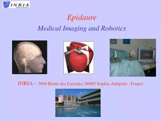
Brain AtlasesEpidaure-LONI Associated teams2002-2004 X. Pennec, N. Ayache, A. Pitiot, V. Arsigny, P. Fillard P. Thompson, A. Toga, J. Annese LONI EPIDAURE Project 2004, route des Lucioles B.P. 93 06902 Sophia Antipolis Cedex (France) Laboratory of Neuro Imaging UCLA California, USA
EPIDAURE LONI Atlas Histology MRI Epidaure LONI Associated teams on Brain Atlases • Functional and structural analysis of the human brain • High variability of the shape of structures • High variability of functions localization • High number of relevant variables • (age, sex, pathologies…) • Building Statistical atlases requires • Powerful algorithms • Large datasets (several hundreds) • Complementarity of the teams • Epidaure: Methodology of image registration/segmentation • LONI: neuroanatomical experts, development/exploitation of large international databases
EPIDAURE EPIDAURE LONI LONI Brain Atlas: Original objectives • Capitalize on the join expertise for more powerful atlases • Algorithms Registration / segmentation tools • Databases MRI, Spect, Histology • Evaluation / validation of the methodologies • Exchange researchers • PhD Co-supervision (A. Pitiot) • Annual visits P. Thompson, A. Toga, N. Ayache, H. Delingette, X. Pennec • Workshop on Brain Atlases summer school on Computational Anatomy
LONI at UCLA • The LONI team TO BE DEVELOPPED • The coordinator: Paul Thompson (To be summurized) • Batchelor 1991, Master 1993, Oxford Univ. • PhD in Neuroscience 1998, UCLA. • Assistant professor at UCLA since 1994 • Publications • 200 refereed publication (1996-2004) (Nature Neuroscience, Nature genetics, TMI, MedIA, J. Neurosciences, NeuroImage…) • Assoc. editor of Human Brain Mapping, Editorial board of MedIA… • Research interests: • Human brain mapping (mathematical and computer intensive methods, building of atlases, encoding variability…) • Brain pathologies (Alzheimer, Schizaphrenia, neuro-oncology) • Barin development (pediatric images)
Quick History • Scientific collaboration initiated in 1999 • Evaluation of histological slices registration (Rat brain images fro LONI) • S. Ourselin et al., “Reconstructing a 3D Structure from Serial Histological Sections”, IVC 2000. • 2 Chapters in “Brain Warping”, A.W. Toga editor, 2003 • J.-P. Thirion, “Diffusing Models and Applications” • G. Subsol, “Crest Lines for Curve Based Warping” • Co-supervision of Alain Pitiot’s PhD (started sept. 2000) • Alain Pitiot, “Segmentation automatique de structures cerebrales s’appuyant sur des connaissances explicites”, Ecole des Mines de Paris, Nov. 2003. • Leveraging by the asociated team program (2002-2003) • 5 peer reviewed publications co-authored by both teams • Software exchanges (10 UCLA publication co-authored by A. Pitiot) • Data exchanges (Arsigny, prize at MICCAI, Media) • New collaborations on brain variability modeling (summer 2003-2005++) • PhD Vincent Arsigny: matching sulcal lines (summer 2003, in preparation) • PhD Pierre Fillard (started sept 2004): article IPMI’05, MICCAI’05
Presentation Overview • To be completed
PhD Alain Pitiot Automatic segmentation of brain structure using explicit knowledge Automated segmentation system maximum a priori knowledge deformable models explicit information Hybrid MRI/histology atlas MRI: in vivo, macroscopic histology: post mortem, microscopic 3-D reconstruction 3-D MRI with superimposed segmented structures 3-D histological volume
Composite segmentation system learnt shape models neural texture filtering piecewise affine registration knowledge-driven segmentation
Neural Texture Filtering Texture classification neural classifier texture maps Hybrid neural architecture hybrid neuralarchitecture classification results
Statistical Shape Modeling Objective statistical shape analysis PCA on shapes Learning approach to reparameterization introduction of explicit knowledge shape distance matrix observed transport shape measure shape modes of variation
Knowledge-driven Segmentation Objective • segmentation of anatomical structures • mix bottom-up constraints with top-down medical knowledge explicit medical information Rules • escape local minima • dynamic control • meta-rules (error checking) segmentation results
Piecewise Affine Registration Motivation • registration of 2-D biological images • adapted transformation model: piecewise affine • specific similarity measure: constrained correlation coefficient Method • similarity map • hierarchical clustering • hybrid elastic/affine interpolation registration results
Joint Contributions wcci-ijcnn ’03 hbm’02 journal of anatomy ipmi’03 hbm’01,’02,’03 neuroimage 2003 wbir’03 tmi 2003 miccai’03 tmi 2002
Executive summary 2002-2003 • Exchange of researchers • PhD Alain Pitiot: joint supervision and localization (50% each) • Visit of N. Ayache and H. Delingette at UCLA (Dec. 2001) • Visit of P. Thompson at Sophia (Nov 2003, PhD defense) • Exchange of software • Reparameterization techniques of Pitiot (IPMI’03)Visual cortex project (Annese HBM’03, Neuroimage 04) • Robust registration software Baladin included in the LONI Pipeline(MAP: Mouse brain Atlas, J. Annese, Visual Cortex, S. Ying, MD, Cerebellum). • Exchange of Data • Neuroanatomical expertise • Segmentations of 4 structures in Brain MR (Pitiot MICCAI’03) • Histological data from J. Annese (Pitiot WBIR’03, Arsigny MICCAI’03) • Segmented Brain sulci (V. Arsigny ++)
Presentation Overview • To be completed
Morphometry of Sucal Lines (summer 2003-2005++) • A unique database • Acquired during 10 years of participation to international projects • More than 500 subjects • 72 manually delineated sulcal lines (roots of grooves on the surface of the brain cortex) • MRI images • Normal and pathological data • Medical annotations • Potentially new anatomical findings
Morphometry of Sucal Lines (summer 2003-2005++) • Innovative methods to model the brain variability • Learn local brain variability from sulci • Learn global correlations in variability and link with asymetry • Better constrain inter-subject registration • Correlate this variability with age, pathologies • Understanding of neurological diseases (Alzheimer, schizophrénie...) • Early diagnosis, follow-up studies
Description fine de la variabilité des lignes sulcales grâce à des techniques d ’analyse innovantes Obtention de nouvelles connaissances anatomiques : corrélations morphologiques entre sillons, asymétrie cérébrale ... Objectifs de l’étude Variance le long des lignes moyennes. Rouge: faible, bleu élevée: Données anatomiques (vert-jaune) etlignes moyennes (rouge) Application : aide au diagnostic, meilleure compréhension de maladies neurologiques (Alzheimer, schizophrénie...) Etiquetage automatique des lignes sulcales
Computation of Average Sulci • Alternate minimization of global variance • Dynamic programming to match the mean to instances • Gradient descent to compute the mean curve position red : mean curve green etyellow : ~80 instances of 72 sulci Sylvius Fissure Arsigny et al. 2004, to appear
Anatomical variability • Variance along the mean sulci • Red (low) to blue (high) Arsigny et al. 2004, to appear
Extraction of Covariance Tensors Currently: 80 instances of 72 sulci About 1250 tensors Covariance Tensors along Sylvius Fissure Color codes Trace Fillard, Pennec, Ayache, Thompson, 2004, to appear
Raw estimation The 4 most representative tensors. Interpolation from the 4 tensors. Compressed Tensor Representation • Mean sulcal line + 4 covariance matrices • optimize for the 4 most representative tensors • Interpolation in-between, extrapolation outside (removes outliers) Sylvian fissure
Compressed Tensor Representation Representative Tensors (250) Original Tensors (~ 1250) Reconstructed Tensors (1250) (Riemannian Interpolation) Fillard-Pennec-Ayache-Thompson 2004, to appear
Variability Tensors Color codes tensor trace Fillard-Pennec-Ayache-Thompson 2004, to appear
Asymmetry Measure Color Codes Distance between “symmetric” tensors
Full Brain extrapolation of the variability Anterior view Color code: principal eigenvector (red: left-right, green: posterior-anterior, blue: inferior-superior) Color code: trace Fillard-Pennec-Thompson- Ayache 2004, to appear
Full Brain extrapolation of the variability Color code: principal eigenvector Color code: trace Fillard-Pennec-Thompson- Ayache 2004, to appear
Scientific results 2004 • Leveraging the Theory • General framework for computing on tensor fields • Interpolation, diffusion, filtering… • “Side results” in Diffusion tensor imaging • Regularization for fiber tracts estimation • Registration,... • Variability of the brain • Learn Variability from Large Group Studies • Statistical Comparisons between Groups • Exploit Variability to Improve Inter-Subject Registration
Executive summary 2004 • Important scientific results • IPAM summer school on Computational Anatomy • 1 week, July 2004, organized by P. Thompson+ 1 week on functional brain imaging • Participation of N. Ayache (Invited speaker), X. Pennec, P. Fillard, and members of Odyssee (O. Faugeras, 2 PhDs). • Tutorial at MICCAI 2004 on evolving processes in Med. Images • Organized by N. Ayache, P. Thompson speaker • Visit of P. Thompson at Sophia afterward
Work plan for 2005 ++ • Modeling the brain variability (PhD P. Fillard) • Validation of the developed models • Learn the full Green’s function Cov(x,y) for all x,y • Theoretical tools already available • Better constrains inter-subject registration (PhD V. Arsigny) • Non stationnarity OK (R. Stefanescu) • Extend to long distnace correlations • Investigate GRID aspects (PhD. T. Glatard) • LONI Pipeline, BIRN • Summer school on Computation Anatomy in Sophia-Antipolis in 2006
Planned budget (2005) • Salaries • PhD Fellowship “complement” for P. Fillard • Visit P. Thompson at Sophia • Missions • Long stay of P. Fillard at LONI A budget is planned at LONI for complementing the salary • Conferences + visits at UCLA: • IPMI’05 (Glenwood Springs, Colorado): X. Pennec + P. Fillard • MICCAI’05 (Palm Springs, CA): N. Ayache, V. Arsigny, X. Pennec
Conclusion • Very active collaboration (software, data, publications) • Access to a unique database (acquision cost > 1 M$) • Potentially new findings in neuro-anatomy and in some brain pathologies