Download
1 / 46
630 likes | 2.37k Vues
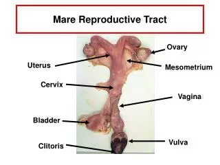
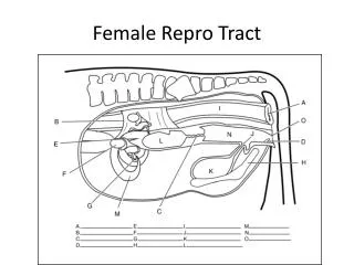
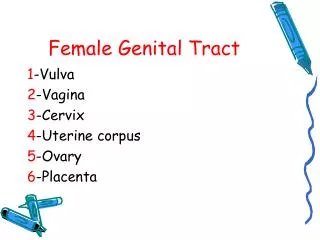
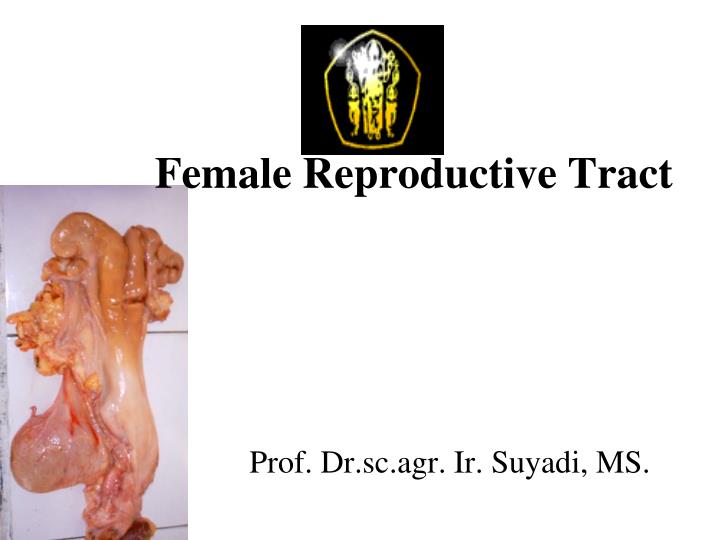
Female Reproductive Tract. Prof. Dr.sc.agr. Ir. Suyadi, MS. Female reproductive tract relative position to the body. Parts of female reproductive organe. Reproductive organs of the female with their major functions. 1. Ovary. Primary reproductive organ in female

E N D
Female Reproductive Tract Prof. Dr.sc.agr. Ir. Suyadi, MS.
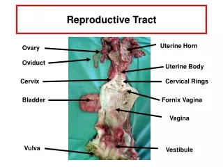
Reproductive organs of the female with their major functions
1. Ovary • Primary reproductive organ in female • Decide monotocous and politocous animals • Almond-shaped, but depend on growing follicles or corpora lutea, in pig is “cluster of grape-like body” • Size (cow): 35x15x15 mm (dep activity) • Consists: • Inner layer medulla: blood vessel, nerves, connective tissue • outer layer surface epithelium (oogonia)
Ovary and Follicle • Beneath the surface epitellium is tunica albuginea ovarii • Below of it is parenchyma • Parenchyma contains follicles and the cells with produce ovarian hormones • Primary follicles are formed during prenatal period • Greatest number at fetal gilt 50 – 90 d; fetal calf 110 – 130 d of gestation
Folllicle: • Located in the parenchyma as in group egg nests, ~ 75,000 primary follicles in young calf. • Continuing follicular growth and maturation 2500 potential ova in old cow. • Some ova are going on maturation and ovulation, while the others undergo to atretic (degenerate).
Follicle: • Are constant in growth and maturation: • Primary follicle: germ cell surrounded by single layer granulosa cells • Secondary follicle by two of more of those cells • Tertiary follicle is characterized by the forming antrum (fluid-filled cavity formed by separating between granulosa cells)
Graafian follicle: • The mature tertiary follicle, which appears as a fluid-filled blister on the surface of the ovary • Fluid in the antrum called liquar folliculi contains steroid reprod hormones, particularly estrogens (:FSH, LH).
Some cell layers in Graafian Follicle: • Theca externa: most outer layer fibrous cells • Theca interna: inside those layer • Base membrane: separates theca interna and innermost layer • Granulosa cells: Innermost cell layer surround antrum cavity • Cumulus oophorus: hillock (mound) of granulosa cells, at one side of the antrum. • Ovum rests upon the cumulus oophorus with other granulosa cells extending around the potential ovum • Corona radiata: grnulosa cells surounding and in immediate contact with ovum.
Cells producing hormones in large follicle: • Theca interna and Granulosa cells are involved in estrogen production • Theca interna cells produces testosterone and converted to estrogen by granulosa cells • Granulosa cells produce progesterone during luteal phase; and some other growth factors (MPF, MK, TGF)
Graafian follicle and ovulation: • The mature follicle will rupture and expellees its contain. This mechanism is occurred under hormonal control. • During ovulation: follicular fluid, some granulosa cells and ovum expelled into body cavity near the oviduct • Oocyte surrounded by corona radiata and other granulosa (cumulus) cells move to the oviduct • The ruptured follicle with blood-filled cavity corpus haemorraghicum • C.haemorgh rapid proliferation of theca externa, theca interna and granulosa cell corpus luteum (yellow body). • Corpus luteum produce progesterone • When CL regresses (in non pregnant animal), it become pale corpus albican
Follicle Corpus haemorragicum
2. Oviduct • Also called fallopian tubes • A pair of convoluted tubes (from near the ovaries to the tips of the uterine horns) • The functions are (see lecture before) • Consists three distinct layers: • The outer layer: tunica serosa (connective tissue) • The middle layer: tunica muscularis (circular and longitudinal smooth muscle fibers) • The innermost layer: tunica mucosa (ciliated and secretory epithelial cells)
Oviduct, morphology • Length: 20 – 30 cm • Divided into 3 segments: • Infundibulum: funnel shape opening near the ovary (mucosal cells are ciliated) • Ampula: middle segment, 3 – 5 cm Ø, half of total length of oviduct; 20 – 40 longitudinal folds • Isthmus: 0,5 – 1 cm Ø, thicker smooth muscle layer than ampula, 4 – 8 mucosal folds; more secretory than ciliated cells. • Ampullary-isthmic-juction: delays the ovum several hours during transport; site of fertilization • Utero-tubal-junction (UTJ): short segment between oviduct and uterus. • Oviduct is stimulated by estrogen and inhibited by progestins
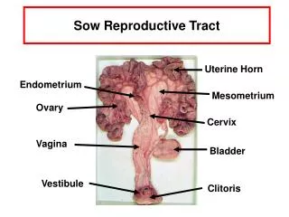
3. Uterus • Extending from UTJ to the cervix • 35 – 60 cm long for cow, sow, mare • Consists: uterine horn and uterine body • Uterine horn: 80 – 90% of total uterine length in cow, sow, doe, ewe; 50% in mare • In ewe and doe, the uteri are less than half of the other above animals
Uterus, function • Major function: retain and nourish of embryo or fetus • Produces uterine milk secreted by glands in the mucosal layer (as nutrients for the embryo before attachment to uterus) • After implantation, placenta plays those important role (nutrients and waste)
Uterus, 4 basic types: • Bicornuate uterus: cow, sow, ewe, doe • Small uterine body, long uterine horn • Fusion of uterine horns biforcatio uteri • Bipartite uterus: mare • Large uterine body and small uterine horns • During pregnancy, fetus develops in both uterine horns but not in uterine body • Duplex uterus: rat, rabbit, guinea pig • Consists two uterine horns each with a seprate cervical canal • Simple uterus: human, primates • A pear-shaped body with no uterine horns
Uterus; histology, • Outer layer tunica serose • Middle layer: myometrium two longitudinal layer of smooth muscle, with a thicker circular layer sandwiched between. Estrogen stimulate to erect myometrium by increasing the tone. Progestins decreases the tone and causing it more flaccid. • Inner layer: endometrium, mucosal lining uterus, is more complex, has simple glands. Progestins cause the endometrial glands to coil and branch, and to secrete uterine milk. The synergistic actions of estrogens and progestins on the endometrium are for preparation of the uterus for pregnancy.
Uterus, physiology • Endometrium provides a mechanism for attachment of the extraembryonic membranes. • This union forms placenta placentation • Type of placenta: see next page
Uterus, Type of Placenta: a. Cotyledonary (cow, ewe, doe): chorionic villi from extraembryonic membranes penetrate into caruncules, which are button-like projection on the endometrium. This union (chorionic villi and caruncle) forms placentome / cotyledon. There are 70 – 120 cotldn attachments in late pregnancy of cow, 88 – 96 in ewes and does. b. Difuse (surface) attachment (sow, mare): Extra embryonic membrane lie in folds on the endometrium, with chorionic villi extending into the endometrium in a more fragile attachment
Uterus, Type of placental attachment: • Epitheliochorial (sow, cow, mare): no erosion in both of endemetrium and extra embryonic membrane during placenta formation (nutrients and maternal blood must pass through both maernal and extra embryonic tissue to reach embryonic blood) • Syndesmochorial (ewe, doe): erosion in the epithelial layer of endometrium • Hemochorial (huma, primates): extension erosion in the epithelial of endometrium (nutrients form maternal blood must pass through only extraembryoic tissue layers to reach fetal blood). • Hemoendothelial (rabbits): no extensive erosion in both extraembryonic membrane and epithelial layer of endometrium
4. cervix • Technically a part of uterus with thick-walled and inelastic; • The anterior being continous with the uterine body; • The posterior end protrudes into the vagina • Length: 5 – 10 cm, 2 – 5 cm, • Contains canal, with opening into uterus • Cervical canal (cow, ewe, doe, sow) transverse interloking ridge : annular rings to seal uterus from contamination.
Cervix; anatomy, histology • In sow, cervical canal is funnel shaped, with ridges having corkscrew configuration • In mare, cervical canal is more open • Histology: • Outer layer: tunica serosa • Middle layer: connective tissue with smooth muscle fibers • Inner layer: mucosa layer contains secretory and ciliated epithelial cells
Cervix, physiology • Estrogen dilatation during estrus and parturation (plus relaxin) • Estrogen epithelial cells mucus protection to microorganisms • During pregnancy, thick mucus – a gel-like plug
5. Vagina • Tubular, thin-waled, quite elastic • 25 – 30 cm long in cow, mare; 10 – 15 cm in sow, doe, ewe • Outer layer – tunica serosa, followed by a smooth muscle layer (circular and longitudinal fibers) • Under progestins influence epithelial lining regenerates.
6. Vulva • External genitalia • Vestibule: common lumen both urinary and reproductive system (10 – 12 cm, cow and mare) • It joins with vagina at urethral orifice • Suburethral diverticulum, just posterior to the external urethra orifice • Labia minora (inner folds / lips); labia majora (outer folds / lips) • Clitoris – gland panis contains erectile tissue and sensory nerves • Vestibular glands, posterior of vestibula. During estrus secrets lubricating mucus
Ovigenesis / oogenesis: • Formation and maturation of the female gamete • Begins in the prenatal period • Following formation, oogonia proliferate by mitotic division within parenchyme • Oocytes enter prophase of the first meiotic division during the fetal period with meiosis I being arrested in late prophase shortly after birth dictyate oocytes (dormant state)
Oogenesis • Full oocyte maturation occurs after puberty • Some oocytes are going to mature and ovulation (<1%), and some others undergo to atresia • Maturation will continue in a cyclic manner after puberty • During each estrous cycle a group of oocytes will start maturation while the other remain dormant. • One of the group that starts development becomes dominant mature ovulation
Principle Maturation stages for the germ cell during ovigenesis • During the fetal period mitosis of oogonia is completed and meiosis I starts • Meiosis I is arrested shortly after birth at prophase I • Growth of oocyte and formation of the zona pellucida are followed closely by growth of the follicle • The preovulatory surge of LH initiates a resumption of meiosis • Meiosis I is completed but meiosis II is arrested at metaphase II • During fertilization, meiosis II resumes and is completed with formation of the zygote.
Oogonium 2N Fetal Period Mitotic division 2N Primary Oocyte • Growth of primary oocyte • Formation of ZP Primary oocyte 2N Zona Pellucida Meiosis I After Puberty Mare Secondary oocyte + 1st polar body N Ovulation Ewe Cow Sow Meiosis II N Zygote + 2nd Polar body
Ovigenesis • Following arrested meiotic development (primary oocyte), maturation resumes with growth of the zona pellucida • Growth of oocyte followed by growth of follicle • FSH stimulates proliferation of granulosa cells: follicle primary secondary formation of antrum • LH theca cells ovulation of dominant follicle • Dominant follicle estrogen preovulatory LH surge • LH oocyte release to antrum fluid (from arrested meiosis I) • Meiosis I produces: 2nd oocyte + 1st polar body : change from 2N N
Oogenesis • Secondary oocyte retain all of cytoplasm and half of nuclear material • Half of nucleus 1st polar body • 1st meiotic div is completed just before ovulation in cow, sow and ewe, and shortly after ovulation in mare
Oogenesis • Second meiotic div (meiosis II) begins immediately after completion of the first division arrest in metaphase II • Meiosis II begins again during fertilization process (will not complete without interaction bwtn oocyte and sperm) • Fertilization: zygote + 2nd polar body
Ovulation • Oocyte + follicle mature LH surge ovulation 24 – 45 h. • LH surges in FF: progesterone Estrogen + PGF2 (inhibition of P, E, PGF block ovulation) • PGF • Ruptures lysosome-like-vesicle: containing proteolytic enzyme (btwn surface epithelium and t. albuginea) t. albuginea, t. externa, t. interna • Actives plasmin: proteolytic enzyme in FF basement membrane
Ovulation • Stigma formed at the apex of follicle, at the point of rupture • Follicle rupture FF, 2nd follicle, granulosa cells are extruded into periotoneal cavity near the infundibulum • Contraction of ovary under PGF control rupture of follicle + expulsion of oocyte • Oocyte surround by cumulus cells cumulus-oocyte-complexes (COC)
Gamete transport: Oocyte • Following ovulaiton: COC is picked up by ciliated epithelial cells of infundibulum • Movement of oocyte to uterus direction: • The cilia cells beat(oocyte and fluid) in the direction of uterus • Segmented, peristaltic contraction of the ampulla • Transport from ampulla to AIJ rapidly, remains at AIJ 2 – 3 days • Cattle : 90 h • Sheep : 72 h • Horse : 98 • Swine : 50
Gamete transport: Spermatozoa • Mechanism: not clear (more speculative) • From deposition site to AIJ : within 8 h • Sperm transport mechanism: • Contraction of cervix, uterus, oviduct • Under stimulation of PGF + estradiol • Sperm motility
Gamete transport: Spermatozoa • Transport barriers: • Cervix : folds + mucus • Random movement of sperm in uterus • UTJ • Lower Isthmus (also as sperm resevoir)