Download
1 / 45
550 likes | 1.1k Vues
NON ACCIDENTAL TRAUMA. Pediatric Critical Care Medicine Emory University Children’s Healthcare of Atlanta. Introduction. >40% Of Death in children <12mos #1 cause of death is head injury 30% of head injury may be misdiagnosed

E N D
NON ACCIDENTAL TRAUMA Pediatric Critical Care Medicine Emory University Children’s Healthcare of Atlanta
Introduction • >40% Of Death in children <12mos • #1 cause of death is head injury • 30% of head injury may be misdiagnosed • 4 of 5 deaths cause by head injury can be prevented if early diagnosis during prior medical evaluation
Epidemiology • Most often < 1 yr of age • Battering is the most common mechanism of injury in children 3-5 mos • Incidence of inflicted TBI is similar in US & Europe
Epidemiology • 60% of cases with previous history or clinical evidence of maltreatment • 22% with involvement of child welfare agencies • 32% with misdiagnosis - Viral gastroenteritis or influenza - “R/O sepsis” - Accidental head injury
Epidemiology • Perpetrators • 50% fathers • 20% step-fathers or male partners • 12% mothers • 17% female baby sitters
Epidemiology • Risk factors • Young/single parents (risk increases more with presence of step-father or maternal boyfriend) • Lower education • Unstable family situation • Stress to family- financial, food & housing, domestic violence, alcohol drug abuse, parental depression • Other: peri-natal illness, family disruption & separation, colicky babies
Mechanism of Injury • Degree of injury in the absence of significant trauma or sign of external injury • Rotational & impact forces • Translational deceleration • Repetitive events – more damage • Developmental weakness: large head, weak & unstable neck; soft brain with higher water contents and poorly demyelinated
Mechanism of Injury • Rotational & Impact forces - Angular deceleration (head rotates on its own axis) causing SDH & axonal injury - > with shaking and impact than shaking alone
Mechanism of Injury • Translational deceleration (drop or short fall) • Head moves in a straight line • Cranial impact • Focal injury
Mechanism of Injury • Significant of cerebral injury is caused by secondary hypoxic ischemic events • Central apnea from injury to the brain stem or cervical spinal cord • Prolonged seizures • Aspiration
Cranial Injury • Blunt force trauma • Shaking • Combination of forces
Shaking Classical pattern • Diffuse unilateral or bilateral SDH • Diffuse multilayered retinal hemorrhage • Diffuse brain injury In the absence of • A history of trauma • Paucity of external manifestation of injury
Intracranial Hemorrhage • Sub-arachnoid • Sub-dural • Intraparenchymal • Epidural
ICH • Short vertical fall <4ft • 85 % with no evidence or minor injury • 7% with skull fracture – all with isolated and linear skull fracture
ICH – Sub-dural hemorrhage • Rare in accidental trauma unless with severe forces (MVA or significant height) • Small and localized to the site of the impact • Interhemispheric SDH usually posterior - 71% of abused children - 19% in accidental injury
ICH – Sub-dural hemorrhage • Mixed density collections of fluid are more common and can present both acute or acute on chronic • Clinical silent SDH • Term infant/neonate with minor birth trauma • Self resolved or increase in size – few days to weeks
ICH – Epidural hemorrhage • Less likely with abuse • More accidental trauma • Focal to the site of impact
ICH – Subarachnoid hemorrhage • Hard to detect • Not good correlation with abuse • Detected mostly at autopsy
ICH – Parenchymal Injury • Contact forces • Inertia forces with rotational deceleration • Traumatic Axonal injury • Sub-cortical white matter, corpus collosum, periventricular regions, dorsolateral aspect of the rostral brainstem • Global Hypoxic Ischemic injury - May cause primary brainstem damage - Prolonged seizure - Secondary hypotension
ICH – Parenchymal Injury • Infarct, atrophy • Encephalomalacia with ventriculomegaly
Associated Injury – Retinal hemorrhage • Numerous • Multi-layered • Extend beyond the posterior pole to the peripheral retina
Associated Injury • Bone fractures • Blunt trauma to abdomen and pelvis
Skull Fractures • Most common parietal • Both accidental & non-accidental • Common sites in abuse • Crossing suture lines • Multiple • Diastatic • Growing • Depressed • Complex • Bilateral
Skeletal Fractures • 20-50% of abused children associated with extracranial skeletal fracture • Ribs, long bone and metaphyseal • Classic metaphyseal avulsion lesion of long bone caused by torsion and traction when extremities in twisted or pulled
Rib Fractures • Most common posterior and lateral • 82% associated with abuse • 8% accidental • 8% bone fragility • 2% birth trauma ** Chest compression more commonly causes lateral and anterior rib fractures
Associated Injuries: Blunt Trauma • Thoracic • Esophageal injury: can result from forced F.B. ingestion, forced caustic ingestion, blunt external trauma, and penetrating trauma • Sx: non specific, pain to the neck and shoulder, shortness of breath, dysphagia, abdominal pain • Early signs: tachycardia, dyspnea, abdominal guarding, pneumothorax, mediastinal air, subcutaneous emphysema
Associated Injuries • Pulmonary Injury • Pulmonary laceration, contusion or diffuse alveolar damage • Chylothorax • Cause by rupture of thoracic duct from blunt trauma or anteroposterior acceleration/deceleration forces • Signs; respiratory distress, nutritional deficiency, electrolytes abnormality, immunosuppression from T-cell depletion
Associated Injuries • Cardiac Injury • Dysrhythmias: commotiao cordis or cardiac concussion causes sudden cardiac arrest (blow at upstroke of the T wave associated with v-fib, blow at the peak of QRS results in asystole • Direct trauma: impact of the heart against the sternum or crushing of the heart due to blunt trauma to the anterior chest • Others: traumatic VSD, cardiac aneurysm, laceration or rupture
Associated Injuries • Abdominal Injury • 1% of abused children suffered intra-abdominal injury with 50% mortality • Sx: tenderness, distension, enlargement of the liver or spleen, and/or bruising of the abdominal wall • Liver injury: most common organ injured; cause contusion, subcapsular hematoma, laceration and rupture • Splenic injury: less common than liver • Pancreatic injury • GI tract • Perforation more common in NAT • Hematoma: intramural hematomas occur most frequently in the duodenum and can cause perforation or stricture
Associated Injuries • Urinary Tract Injury • Renal injury: contusion or subcapsular hematoma, shattered kidney or vasculaar pedicle avulsion • Hematuria is present in 41-68% of victims with renal trauma • Ureteral injury • Bladder injury: bladder rupture (blunt force to a full bladder). Rupture occurs at the dome of the bladder, fluid and blood extravasate into the peritoneum
Evaluation • History • Physical Examination • Laboratory studies • Radiographic studies
Evaluation: History • Who, what, when and where • Document your history • Document inconsistency of the story through details • Help your memory at a later time (across a DA and a defense lawyer)
Evaluation: History • Who was present? • Who had been taking care of the patient at least 4 hours prior to the event • When did the last time the child seem normal? When was the event • Review the event after the child last seen to be normal
Evaluation: History • Where did the event occur? Who was there with the baby? • What would care provider consider normalcy in the patient? (behavior, development)
Evaluation: History • Don’t forget details of family history • Bleeding tendency in family • Bleeding at time of circumcision for boys • Easy bruising
Evaluation: Laboratory • CBC with Platelet • Coagulation study: DIC panel • Electrolytes, liver function test, and urinalysis • * preliminary evidence of CSF and serum measuremenf of biomarkesr of brain injury – neuron-specifiec enolase, S100B(a calcium binding protein found in astrocytes), and myelin basic protein
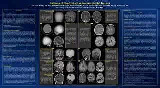
Evaluation: Imaging • CT – brain and bone window is best as an initial tool. • MRI – superior to CT for documenting the pattern, extent, and timing • Skeletal survey
Evaluation: Opthalmologic Exam • Need to have an opthalmologic exam to stand legally
Differential Diagnosis • Accidental injury • Birth trauma • Apparent Life-threatening event • Bleeding disorder • Others
Differential Diagnosis – Accidental Injury • A history of traumatic event • Retinal hemorrhages are typically fewer in number and less extensive • Subdural hematomas
Differential Diagnosis – Birth Trauma • Commonly associated with instrumented deliveries • Both retinal hemorrhage and subdural hemorrhage
Differential Diagnosis – Bleeding Disorder • ICH can occur in severe bleeding disorer (hemophilia) spontaneously or following an injury • Retinal hemorrhages are small in number and are typically confined to the posterior pole • Boys with hemophilia, ICU occurs most often in the neonatal period • ICH is uncommon in idiopathic thrombocytopenic purpura