Download
1 / 50
1.88k likes | 5.51k Vues
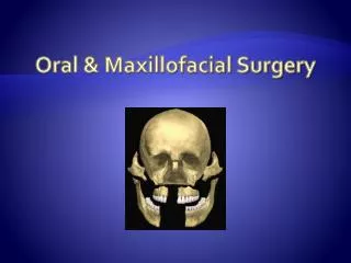
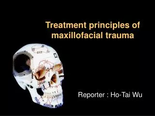
Maxillofacial Trauma. Anatomy. Anatomy. Maxillofacial Region. 1. Fractures of the Nasal Pyramid 2. Fractures of the Central Midface Le Fort Fractures. Maxillofacial Region. 3. Fractures of the Lateral Midface 4. Fractures of the Frontal bone 5. Fractures of the Anterior Skull Base

E N D
Maxillofacial Region 1. Fractures of the Nasal Pyramid 2. Fractures of the Central Midface • Le Fort Fractures
Maxillofacial Region 3. Fractures of the Lateral Midface 4. Fractures of the Frontal bone 5. Fractures of the Anterior Skull Base • Escher Classification
Maxillofacial Region 6. Fractures or dislocation of the mandible
Etiology • Sports • Vehicular Accidents • Mauling • Women – consider the possibility of domestic violence
Etiology • Patients with severe facial trauma: • multisystem trauma • potential for airway compromise • concurrent brain injury • cervical spine injuries • blindness
Emergent Management • Primary Survey • Airway • Breathing • Circulation • Secondary Survey
Emergent Management Airway: • Chin lift. • Jaw thrust. • Oropharyngeal suctioning • Manually move the tongue forward • Maintain cervical immobilization
Emergent Management • Avoid nasotracheal intubation • Adverse effects: • Nasocranial intubation • Nasal hemorrhage • cricothyroidotomy
Emergent Management Circulation: • Direct pressure • Anterior and posterior nasal packing • Packing of the pharynx around ET tube
History • Place, Time, Date, Mechanism of injury • Detailed description of the circumstances surrounding the injury • Allergies, other medical problems, medications, tetanus immunizations
History • Questions: • Was there LOC, nausea/vomiting, headache? (Head Trauma related questions) • How is your vision? • Hearing problems? • Is there pain with eye movement? • Are there areas of numbness or tingling on your face? • Able to bite down without any pain? • Is there pain with moving the jaw?
Physical Examination Inspection • Open wounds for foreign bodies • Facial asymmetry • Nose for deviation, widening of bridge • Nasal septum for septalhematoma, CSF or blood • Ears for blood or CSF • Malocclusion
Physical Examination Inspection • Raccoon eyes • Battle’s sign
Physical Examination Inspection • Halo Sign • Not sensitive or specific but can be used as a preliminary test for CSF in blood • Dipstick • Beta transferrin • Otorrhea, Rhinorrhea
Physical Examination Palpation • Palpate the entire face. • Supraorbital and Infraorbital rim • Zygomatic-frontal suture • Zygomatic arches • Nose - crepitus, deformity and subcutaneous air • Zygoma along its arch and its articulations with the maxilla, frontal and temporal bone • Mandible for tenderness, swelling
Physical Examination • Intraoral examination: • Inspect the teeth for malocclusions, bleeding • Manipulation of each tooth • Check for lacerations • Mandibular movements
Physical Examination Ophthalmologic exam • Visual acuity • Pupils for shape and reactivity • Eyelids for lacerations • Extra ocular muscles • Palpate around the orbits
Physical Examination • Examine and palpate the exterior ears • Otoscopic examination • Look for lacerations • TM rupture
Diagnostic Imaging • Plain films • Confirm suspected clinical diagnosis • Determine extent of injury • Document fractures • CT scan
General Treatment • ATS, TeAna • Thorough evaluation of all wounds • All foreign bodies must be removed • Debridement • Suturing of lacerations as needed • Minimize scarring • Antibiotics
Nasal Fractures • Most common bone injury in the face • Open or closed • Signs • Depression or displacement of nasal bones • Edema of nose • Epistaxis • Fracture of septal cartilage with displacement or mobility • Crepitus on palpation
Nasal Fractures • All nasal injuries should be evaluated for septal hematoma • Untreated- result in septal necrosis and saddle nose deformity • Can become infected- result in a septal abscess
Nasal Fractures • Radiographs: • Lateral projection • Treatment: • Surgical • After reduction, nasal cavities should be packed – “internal splinting”
Maxillary Fractures • Le Fort’s classification • Le Fort I (transverse maxillary) • Le Fort II (pyramidal) • Le Fort III (craniofacial dysjunction)
Le Fort I • Low transverse fracture of maxilla involving palate • Facial edema • Mobility of hard palate and upper teeth • Malocclusion
Le Fort II • Pyramidal fracture with detachment of maxilla • Facial edema • Epistaxis • Bilateral periorbital edema and ecchymosis
Le Fort III • Complete disruption of attachments of facial skeleton to cranium • Movement of all facial bones in relation to the cranial base with manipulation of the teeth and hard palate • Open patient’s mouth and grasp the maxilla arch • Place the other hand on the forehead • Gently move back and forth, up and down - check for movement of maxilla
Le Fort III • Massive edema with facial elongation, flattening – “Dish faced deformity” • Epistaxis and CSF rhinorrhea • Motion of the maxilla, nasal bones and zygoma
Management of Le Fort Fractures • Open reduction and intermaxillary fixation should be performed to establish correct occlusion • Followed by rigid fixation at the piriform rims and zygomaticomaxillary buttress.
Zygoma Fractures • The zygoma has 2 major components: • Zygomatic arch • Zygomatic body • Two types of fractures can occur: • Isolated Arch fracture -most common • Tripod fracture - most serious
Zygoma Arch Fractures • Palpable bony defect over the arch • Flattening of the cheek • Pain in cheek and jaw movement • Limited mandibular movement
Zygoma Arch Fractures • Radiographic imaging: • Submental view “bucket handle view” - Arches may not be seen in usual views (anterior, lateral) • Treatment: • Symptomatic - surgical
Zygoma Tripod Fractures • Tripod fractures consist of fractures through: • Zygomatic arch • Zygomaticofrontal suture • Inferior orbital rim and floor • Symptoms • Periorbital edema • Sensory disturbances along the infraorbital nerve
Zygoma Tripod Fractures • Waters • Caldwell • Submental • Coronal CT • Treatment: • Symptomatic - surgical
Orbital Blow Out Fractures • Isolated fracture of the orbital floor with partial herniation of orbital contents • Facial asymmetry • Enophthalmos • Diplopia on upward gaze- impingement of inf. Rectus • Check for sensory disturbances – cheek, upper lip, lateral nasal wall
Orbital Blow Out Fractures • CT scan • Management: • Indicated for displaced fractures or for symptomatic fractures
Frontal Sinus Fracture • Uncommon • Depression of anterior table of frontal sinus • Intracranial injuries • Dural tears • Epistaxis • CSF rhinorrhea (disruption of posterior table of frontal sinus with dural rupture)
Frontal Sinus Fracture • Radiographs: • Facial views should include: • Waters • Caldwell • lateral projections • Caldwell view best evaluates the anterior wall fractures
Frontal Sinus Fractures • Cranial CT with bone window • Frontal sinus fractures. • Orbital rim and nasoethmoidalfractures • R/O brain injuries or intracranial bleeds
Frontal Sinus Fractures • Patients with depressed skull fractures or with posterior wall involvement. • ENT or nuerosurgery consultation. • Admission. • IV antibiotics. • Tetanus. • Patients with isolated anterior wall fractures, nondisplaced fractures can be treated outpatient after consultation with neurosurgery.
Frontal Sinus Fractures • Associated with intracranial injuries • Orbital roof fractures • Dural tears • Mucopyocoele • Epidural empyema • CSF leaks • Meningitis
Mandibular Fractures • 2nd most commonly fractured facial bone • Signs and symptoms • Malocclusion of teeth • Tooth mobility • Intraoral lacerations • Pain on mastication • Bone deformity
Mandibular Fractures • Mandibularpain • Malocclusion of the teeth • Separation of teeth with intraoral bleeding • Inability to fully open mouth • Preauricular pain with biting • Positive tongue blade test
MandibularFractures • Radiographs: • Panorex • Plain view: PA, Lateral and a Townes view
MandibularFractures Treatment: • Nondisplacedfractures: • Analgesics • Soft diet • Dent/ORL surgery referral • Displaced fractures, open fractures and fractures with associated dental trauma • Urgent oral surgery consultation • All fractures should be treated with antibiotics and tetanus prophylaxis.