Download
1 / 45
490 likes | 900 Vues
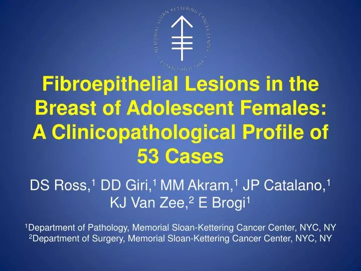
Fibroepithelial Lesions in the Breast of Adolescent Females: A Clinicopathological Profile of 53 Cases. DS Ross, 1 DD Giri, 1 MM Akram, 1 JP Catalano, 1 KJ Van Zee, 2 E Brogi 1 1 Department of Pathology, Memorial Sloan-Kettering Cancer Center, NYC, NY

E N D
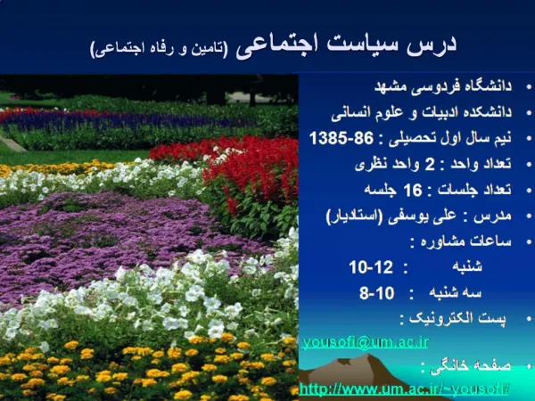
Fibroepithelial Lesions in the Breast of Adolescent Females:A Clinicopathological Profile of 53 Cases DS Ross,1 DD Giri,1 MM Akram,1 JP Catalano,1 KJ Van Zee,2 E Brogi1 1Department of Pathology, Memorial Sloan-Kettering Cancer Center, NYC, NY 2Department of Surgery, Memorial Sloan-Kettering Cancer Center, NYC, NY
Background Fibroepithelial lesions (FELs): • Most common breast abnormality in adolescent females (< 18 years-old) • Referred to as: • Fetal fibroadenoma • Cellular fibroadenoma • Juvenile fibroadenoma • Giant fibroadenoma • Fibroadenoma variant • Tubular adenoma • Hamartoma • Cystosarcoma phyllodes • Phyllodes tumor
Study Aims • Investigate the morphology and clinical behaviorof FELs in adolescents • Standardize the diagnostic terms used for FELs in adolescents based upon histologic criteria
Study Design (1) • Search of MSKCC pathology database • Age < 18 years-old • Excision or mastectomy between 1992-2011 • Diagnosis of any fibroepithelial lesion • 7 FELs from a prior series were also included (1975-1983) Barrio AV et al. Ann Surg Oncol 2007
Study Design (2) • 3 pathologists reviewed all available slides • Features noted: • Gross & microscopic size • Borders / margin status • Growth pattern • Stromal overgrowth • Stromal cellularity • Nuclear pleomorphism • Epithelial hyperplasia • Stromal cell mitoses • Smooth muscle actin-α (1A4, DAKO) staining performed on available tissue
Study Design (3) • Patient information and clinical follow-up obtained from e-medical records • Age at presentation • Age at menarche • Race • Laterality • Family history
Patient Population (1) *Information available for 26 patients **1 patient presented 12 mo prior to menarche
Patient Population (2) *Pt undergoing US for ipsilateral FEL
Malignant Phyllodes Tumor (8%), Low Grade N=1,High Grade N=3 • Gross size*: 4 & 25cm • Infiltrative borders** • Stromal overgrowth • All high grade tumors *Gross size available for 2 cases **Borders assessable in 2 case
Malignant Phyllodes Tumor (8%), Low Grade N=1,High Grade N=3 • Increased stromal cellularity • Moderate-marked stromal nuclear atypia • Mean mitotic count / 10 HPF: • Low grade: 10 • High grade: 17 (12-20)
Benign Phyllodes Tumor, N=15 (28%) • Mean gross size*: 4 cm (1-13 cm) • Borders**: • Circumscribed: 11/14 (79%) • Infiltrative: 3/14 (21%) • Stromal overgrowth: 13% (2/15) • Growth pattern • Intracanalicular: 10/15 (67%) • Pericanalicular: 5/15 (33%) *Gross size available for 11 cases **Borders assessable in 14 cases
Benign Phyllodes Tumor, N=15 (28%) • Increased stromal cellularity • Mild-moderate stromal nuclear atypia • Epithelial hyperplasia: 7/15 (47%) • Mean mitotic count / 10 HPF: 3.1 (1-7)
Benign Phyllodes Tumor, N=15 (28%) • Increased stromal cellularity • Mild-moderate stromal nuclear atypia • Epithelial hyperplasia: 7/15 (47%) • Mean mitotic count / 10 HPF: 3.1 (1-7)
Benign Phyllodes Tumor, N=15 (28%) • Increased stromal cellularity • Mild-moderate stromal nuclear atypia • Epithelial hyperplasia: 7/15(47%) • Mean mitotic count / 10 HPF: 3.1 (1-7)
Usual Fibroadenoma, N=11 (21%) • Mean gross size: 2.6 cm (0.7-4.5 cm) • Circumscribed borders • Growth pattern • Intracanalicular: 10/11 (91%) • Pericanalicular: 1/11 (9%)
Usual Fibroadenoma, N=11 (21%) • Increased stromal cellularity: 3/11(27%) • No stromal nuclear atypia • Epithelial hyperplasia: 2/11(18%) • Mean mitotic count / 10 HPF: 1.3 (0-6) • 6 mit / 10 HPF in pt pregnant 1y before; FA w/ focal lactational changes • Mean mitotic count w/o above pt is 0.8
Usual Fibroadenoma, N=11 (21%) • Increased stromal cellularity: 3/11 (27%) • No stromal nuclear atypia • Epithelial hyperplasia: 2/11(18%) • Mean mitotic count / 10 HPF: 1.3 (0-6) • 6 mit / 10 HPF in pt pregnant 1y before; FA w/ focal lactational changes • Mean mitotic count w/o above pt is 0.8
Usual Fibroadenoma, N=11 (21%) • Increased stromal cellularity: 3/11 (27%) • No stromal nuclear atypia • Epithelial hyperplasia: 2/11 (18%) • Mean mitotic count / 10 HPF: 1.3 (0-6) • 6 mit / 10 HPF in pt pregnant 1y before; FA w/ focal lactational changes • Mean mitotic count w/o above pt is 0.8
Juvenile Fibroadenoma, N=23 (43%) • Mean gross size*: 3.1 cm (0.5-7 cm) • Circumscribed borders • Growth pattern • Pericanalicular • Floridly glandular • Retention of lobular structure *Gross size available for 22 cases
Juvenile Fibroadenoma, N=23 (43%) • Increased stromal cellularity: 14/23 (61%) • No stromal nuclear atypia • Epithelial hyperplasia: 7/23 (30%) • Mean mitotic count / 10 HPF: 1.8 (0-7)
Juvenile Fibroadenoma, N=23 (43%) • Increased stromal cellularity: 14/23 (61%) • No stromal nuclear atypia • Epithelial hyperplasia: 7/23 (30%) • Mean mitotic count / 10 HPF: 1.8 (0-7)
Juvenile Fibroadenoma, N=23 (43%) • Increased stromal cellularity: 14/23 (61%) • No stromal nuclear atypia • Epithelial hyperplasia: 7/23(30%) • Mean mitotic count / 10 HPF: 1.8 (0-7)
Juvenile Fibroadenoma • A distinctive type of FA • Collagenized and cellular stroma • Pericanalicular growth pattern • Lobular arrangement • +/- Florid ductal hyperplasia
Juvenile Fibroadenoma: Variation in Morphology
Fibroadenoma Variant • Characterized by collagenous and cellular stroma • “…related to FAs in structure and composition but show sufficient difference to raise problems in precise dx and classification” • Differential dx often includes a phyllodes tumor Azzopardi, 1979
Juvenile Fibroadenoma, Variant Pattern (N=8/23) Problems in Breast Pathology, Azzopardi, 1979 Case from our series
Juvenile Fibroadenoma: Variant Pattern & Intratumoral Heterogeneity (N=4/23)
Juvenile Fibroadenoma: Variant Pattern & Intratumoral Heterogeneity (N=4/23)
Distinguishing Features Juvenile Fibroadenoma Benign Phyllodes Tumor
Distinguishing Features Juvenile Fibroadenoma Benign Phyllodes Tumor
Distinguishing Features Juvenile Fibroadenoma Benign Phyllodes Tumor Mean Mitotic Count: 1.8 (0-7) Mean Mitotic Count: 3.1 (1-7)
Follow-Up • 37 patients (41 FELs) • Mean follow-up: 40 months • From less than a month to 278 months
2 Recurrent Cases • Information for index FEL
Conclusions • FEL in adolescents • Benign in two-thirds of cases • Juvenile FA 46%, Usual FA 24%
Conclusions • FEL in adolescents • Benign in two-thirds of cases • Juvenile FA 46%, Usual FA 24% • Frequent stromal mitoses
Conclusions • FEL in adolescents • Benign in two-thirds of cases • Juvenile FA 46%, Usual FA 24% • Frequent stromal mitoses • Juvenile FA is the most common FEL • Distinctive morphology
Conclusions • FEL in adolescents • Benign in two-thirds of cases • Juvenile FA 46%, Usual FA 24% • Frequent stromal mitoses • Juvenile FA is the most common FEL • Distinctive morphology • +/- Stromal expansion and intratumoral heterogeneity (fibroadenoma variant)
References • Amerson, J.R., Cystosarcoma phyllodes in adolescent females. A report of seven patients. Ann Surg, 1970. 171(6): p. 849-56. • Ashikari, R., J.H. Farrow, and J. O'Hara, Fibroadenomas in the breast of juveniles. Surg Gynecol Obstet, 1971. 132(2): p. 259-62. • Azzopardi, J.G., A. Ahmed, and R.R. Millis, Problems in breast pathology. Major problems in pathology1979, Phildelphia: Saunders. xvi, 466 p. • Barrio, A.V., et al., Clinicopathologic features and long-term outcomes of 293 phyllodes tumors of the breast. Ann Surg Oncol, 2007. 14(10): p. 2961-70. • Ewing, J., Neoplastic Diseases, 2nd ed.1927, Philidelphia: W. B. Saunders. • Hertel, B.F., C. Zaloudek, and R.L. Kempson, Breast adenomas. Cancer, 1976. 37(6): p. 2891-905. • Koerner, F.C.. Diagnostic Problems in Breast Pathology. 2009, Philadelphia: Saunders. • Mies, C. and P.P. Rosen, Juvenile fibroadenoma with atypical epithelial hyperplasia. Am J Surg Pathol, 1987. 11(3): p. 184-90. • Pike, A.M. and H.A. Oberman, Juvenile (cellular) adenofibromas. A clinicopathologic study. Am J Surg Pathol, 1985. 9(10): p. 730-6. • Rosen, P.P., Rosen's breast pathology. 3rd ed 2008, Philadelphia: Lippincott Williams & Wilkins. • Tavassoli, F.A., Pathology of the breast. 2nd ed1999, Stamford, Conn.: Appleton & Lange. xi, 874 • Wulsin, J.H., Large breast tumors in adolescent females. Ann Surg, 1960. 152: p. 151-9.