Download
1 / 25
590 likes | 1.51k Vues
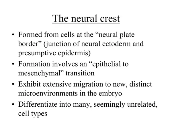
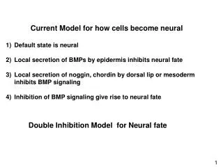
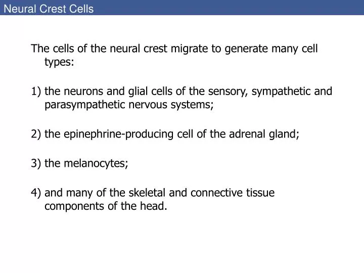
Neural Crest Cells. The cells of the neural crest migrate to generate many cell types: the neurons and glial cells of the sensory, sympathetic and parasympathetic nervous systems; the epinephrine-producing cell of the adrenal gland; the melanocytes;

E N D
Neural Crest Cells • The cells of the neural crest migrate to generate many cell types: • the neurons and glial cells of the sensory, sympathetic and parasympathetic nervous systems; • the epinephrine-producing cell of the adrenal gland; • the melanocytes; • and many of the skeletal and connective tissue components of the head.
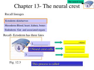
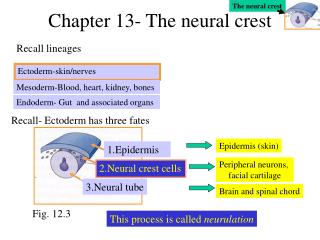
13.1 Schematic representation of neural crest formation in an amniote embryo, cross section (1)
13.1 Schematic representation of neural crest formation in an amniote embryo, cross section (2)
13.3 Neural crest cell migration in the trunk of the chick embryo (Part 1)
13.3 Neural crest cell migration in the trunk of the chick embryo (Part 2) 2-day chick embryo stained red with antibody to HNK-1 which selectively recognizes NC cells. Extensive staining is seen in the anterior part of each sclerotome.
13.3 Neural crest cell migration in the trunk of the chick embryo (Part 3) Cross sections through the chick embryo showing extensive migration through the anterior portion of the sclerotome (C), but (D) no migration through the posterior portion.
13.4 All migrating neural crest cells are stained red by antibody to HNK-1 • What signals initiate migration? • When does the migratory agent become competent to respond to these signals? • How do the migratory agents know the route to travel? • What signals indicate that the destination has been reached? Wnt and FGF BMP4 and BMP7 Slug and RhoB
13.5 Segmental restriction of neural crest cells and motor neurons by the ephrin proteins of the sclerotome (Part 1)
13.5 Segmental restriction of neural crest cells and motor neurons by the ephrin proteins of the sclerotome
13.6 Hair follicle of a mouse Some of these melanocytes migrate outside the bulge to differentiate into mature melanocytes and provide pigmentation to the hair shaft.
13.8 Paracrine factors encountered in the environment help specify the different neural crest-derived lineages in the trunk
13.9 Cranial neural crest cell migration in the mammalian head (Part 1)
13.9 Cranial neural crest cell migration in the mammalian head (Part 2) Contributes to the forehead, nose, philtrum of the upper lip, and to the primary palate. Generates the sides of the nose. Lower and upper jaw, and to the sides of the middle and lower regions of the face.
13.9 Cranial neural crest cell migration in the mammalian head (Part 3)
13.12 Intramembranous ossification (Part 2) BMPs CBFA1 Osteocalcin Osteoporin and others Blue: cartilage Red: bone
13.13 Cranial neural crest cells in embryonic mice, stained for -galactosidase expression (Part 1) A, B, C: day 6-9.5
13.13 Cranial neural crest cells in embryonic mice, stained for -galactosidase expression (Part 2)
13.13 Cranial neural crest cells in embryonic mice, stained for -galactosidase expression (Part 3)
13.14 The septa that separate the truncus arteriosus into the pulmonary artery and aorta form from cells of the cardiac neural crest (Part 1)
13.14 The septa that separate the truncus arteriosus into the pulmonary artery and aorta form from cells of the cardiac neural crest (Part 2)