Download
1 / 19
730 likes | 3.19k Vues
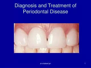
RADIOGRAPHIC DIAGNOSIS OF PERIODONTAL DISEASES. Dr Mohammed Malik Afroz. FORMAT. INTRODUCTION CLASSIFICATION OF PERIODONTAL DISEASES RADIOGRAPHIC TECHNIQUES FOR PERIODONTAL DISEASES RADIOGRAPHIC ASSESSMENT OF PERIODONTAL DISEASES DIGITAL RADIOGRAPHIC ASSESSMENT LIMITATIONS OF RADIOGRAPHS

E N D
RADIOGRAPHIC DIAGNOSIS OF PERIODONTAL DISEASES Dr Mohammed MalikAfroz
FORMAT • INTRODUCTION • CLASSIFICATION OF PERIODONTAL DISEASES • RADIOGRAPHIC TECHNIQUES FOR PERIODONTAL DISEASES • RADIOGRAPHIC ASSESSMENT OF PERIODONTAL DISEASES • DIGITAL RADIOGRAPHIC ASSESSMENT • LIMITATIONS OF RADIOGRAPHS • CONCLUSION
INTRODUCTION • Clinical data is the primary aspect in diagnosing a periodontal condition. • Radiographs act as an adjunct (addition) to the clinical examination • Most cases clinical assessment cannot give a clear picture and hence radiographs are a must. • Clinical examination comprises of – • Gingival architecture • Erythema • Edema • Probing depths • Bleeding indices
Radiographs are essential in terms of • Amount of bone present • Condition of alveolar crest • Bone loss in furcation area • Width of PDL • The structure of lamina dura • Root length and morphology • Crown to root ratio • Anatomic issues: • Maxillary sinus • Missing, supernumerary and impacted teeth • Contributing factors • Caries • Apical inflammatory lesions • Root resorption
Radiographic classification of periodontal disease • Bone Level Assessment • Mild periodontitis • Moderate periodontitis • Severe /Aggressive periodontitis • Furcation Involvement
Radiographic Techniques For Periodontal Examination • Paralleling Cone IOPA radiographic Technique • Most preferred technique as it has least magnification, clear picture for bone presence calculation. • A full mouth IOPA radiographs are taken with this technique and individually assessed for calculation and examination. • Pre and Post operative radiographs are taken for assessment
Radiographic Techniques contd… • Panoramic Radiograph – newer machines have various settings which allows for a least magnified radiograph. • They are able to take all dentition in one radiograph without any magnification • The panoramic machine software is able to calibrate/ calculate the level of bone.
Incipient Periodontitis/ Mild Periodontitis • Is a clinical condition • Can be assessed by gingival and periodontal examination. • Periodontal examination should include using periodontal probe and other diagnostic aids
Moderate Periodontitis • Has bone loss, followed by loss of contour of the alveolar architecture. • There is no presence of furcation involvement. • The level of bone is seen below the CEJ.
Severe/Aggressive Periodontitis • There is bone loss which may range from mild to bone loss till apical 1/3rd or more • There is furcation involvement with one or more teeth. • Bone loss can be angular or vertical or horizontal. • In severe bone loss there is generalized vertical bone loss with most teeth. • May have angular or vertical bone loss in some teeth.
Furcation Involvement • There are three grades of furcation involvement • Grade 1 – seen on radiograph as a triangular radiolucency which is very mild range from 1mm to 3mm • Grade 2 – is seen as a radiolucency having a triangular appearace which is between 3mm to 5mm. • Grade 3 – is a frank triangular radiolucency of more than 5mm.
Modern Radiography for Periodontal Examination • Digital Radiography • Subtraction Radiography • Digital radiography id preferred today as the images are instant and easy to store. • A full mouth digital IOPA radiographs can be stored with the patient file and pre and post operative radiographs can be assessed • Digital radiographs also allow software related calcualation of the bone assessment
Subtraction Radiography • This is more easily applied with the digital radiograph where the pre and post operative radiographs can be assessed together after the digital software cancels the common image and shows only the new image • The calculation for whether really the bone has increased in height can be assessed
Quantitative And Computer Generated Interpretation • Changes of bone density can be measured by means of computer aided procedures. • By use of a calibration wedge with known radiation attenuation properties, the measurements of density differences can be converted into estimations of volume changes. • Aluminum or hydroxyapatite is often used because of its similarity to bone in terms of radiation attenuation characteristics.
Three Dimensional Imaging • Location techniques • Computed tomography • Magnetic resonance imaging • 3 – D Reconstruction
Chance Findings • Most of the time the radiographs show presence of calculus. • It gives a false appearance of extra enamel deposition on the radiograph. • The calculus can be differentiated from enamel based on the density of calculus which is less than enamel • The calculus has a more triangular radiopaque appearance usually seen near the CEJ and the radiodensity merges with the adjacent radiolucency. • Plaque cannot be seen on the radiograph
LIMITATIONS OF RADIOGRAPH • Conventional radiographs provide a two dimensional image of complex, three dimensional anatomy. • Due to superimposition, the details of the bony architecture may be lost • Radiographs do not demonstrate incipient disease, as a minimum of 55- 60%demineralization OF BONE must occur before radiographic changes are apparent • 30% TOOTH DEMINERALIZATION should occur before seen on bone. • Do not record soft tissue contours • Gingivitis is no seen on radiograph
Conclusion • Radiographs are seen as best adjuncts to the clinical examination for periodontal examination • Though it has its limiotations but it always gives a best picture about the condition
Thank You • Any Questions????