Download
1 / 84
980 likes | 2.07k Vues
Congenital LVOT Obstruction. Seoul National University Hospital Department of Thoracic & Cardiovascular Surgery. Congenital LVOT Obstruction. Types of obstruction Supravalvular aortic stenosis Valvular aortic stenosis Subvalvular aortic stenosis Intraventricular obstruction

E N D
Congenital LVOT Obstruction Seoul National University Hospital Department of Thoracic & Cardiovascular Surgery
Congenital LVOT Obstruction • Types of obstruction • Supravalvular aortic stenosis • Valvular aortic stenosis • Subvalvular aortic stenosis • Intraventricular obstruction • Hypoplastic left heart syndrome
Left Ventricular Outflow Tract • Congenital malformations • Obstruction • * Supravalvular • * Valvular • * Subvalvular • * Intraventricular obstruction • - Occurs in combination with other cardiac lesions • ( Interruption, COA, MV apparatus anomalies, • left ventricular hypoplasia ) • Regurgitation • * Annular aortic root dilatation • * Prolapse of valve leaflets • * Degenerative abnormality • * Rupture of aneurysm of sinus of Valsalva
LVOT Obstruction • Pathophysiology • Left ventricular outflow tract obstruction(LVOTO ) leads to left ventricular hypertrophy, ischemia, and ventricular dysfunction. • The obstruction is at the valvar , subvavar, or supravalvar level.
Congenital Aortic Valve Diseases • Manifestation • Incidence • * 2 - 6% of CHD ( about 5%) • * AS : common, M : F = 4 : 1 • * AR : less common, no sex predilection • Etiology • 1) AS : not known or no evidence • # probably genetic aberration in IHS, • and supravalvular stenosis • # fetal aortic valve endocarditis • 2) AR : caused by any one of several disease (rheumatic • fever, endocarditis, Marfan syndrome, Ehlers - • Danlos syndrome, connective tissue disorders)
Congenital Aortic Valve Diseases • Reparative procedures • Aortic stenosis • * Not curative, but palliative • * High mortality in neonate • * Reasonable mortality in infant and children • * Residual stenosis & induced aortic regurgitation • * Overall 10-year survival : 80 - 90% • * 10-year reoperation-free survival : 50 - 60% • Aortic regurgitation • * Medical treatment if possible • * Valvoplasty for prolapsing cusp • * Aortic valve replacement
Congenital Aortic Stenosis • Definition • A cardiac anomaly in which narrowing at valvar, subvalvar, supravalvar, or combined levels results in a systolic pressure gradient between the inflow portion of left ventricle & aorta beyond obstruction. • Classification refersto the predominant area of obstruction in the left ventricularoutflow tract, inevitably,these groups sometimes overlap because of the complexity of pathologicchanges
LV Outflow Tract • Structures
Aortic Stenosis • Types
Aortic Outflow Obstruction • Clinical features • Infantile • 1) Usually appears within the 1st. month of life • 2) Presentation in later infancy according to the severity and growth • 3) Untreated mortality ; 23% in the 1st. year • Childhood • 1) Progressive with growth, rare in early childhood • 2) If left ventricular failure develops, rapidly deteriorate • 3) Sudden death : 1-19%, but rare in low pressure gradient • * Consequence of low aortic pressure (coronary insufficiency) • * Arrhythmia • * Frequent when resting pressure gradient more than 50mmHg • 4) Untreated mortality • * 60% at 40 years • * Mean age of death : 35 years
Aortic Outflow Obstruction • Operation • Indications • 1) Critical AS in neonate ; urgent • 2) Infant and children • * Pressure gradient over 70mmHg • * Sx. of angina, syncope, exercise intolerance, LVH, with pr. gradient • over 50mmHg and valve area less than 0.5 square cm/BSA • * Pressure gradient over 40mmHg in subvalvular lesion • Methods • 1) Valvotomy • * Open & closed technique (hypothermia) • * Balloon valvotomy • 2) Resection of subvalvular tissue & myocardium • 3) Aortoplasty of supravalvular stenosis • 4) Aortoventriculoplasty in tunnel stenosis • 5) Valve replacement
Congenital Valvar Aortic Stenosis • Definition • An obstruction at valve level caused by imperfect • cusp development with leaflet thickening and fusion • History • Marquis, Logan : Surgical treatment by dilator in 1955 • Swan, Lewis : Open valvotomy in 1956 • Spencer : Valvotomy through OHS in 1958
Congenital Valvar Aortic Stenosis • Manifestation • Etiology : unknown • * Malabsorption of conal element ( leaflet dysplasia as in PS) • * Histologic disorganization of aortic media and dysplasia in left • ventricular septum (Somerville) & hypoplasia of annulus rarely • Incidence : 3~6% of all CHD, 60 - 70% of AS • Anatomy • * Hypoplasia of annulus : rare • * Abnormally formed valve leaflets : majority • Bicuspid ; 70% (left and right) • Unicuspid • Thick and dysplastic valve • - Commonly associated with COA, MV abnormalities, sub or • supravalvar stenosis, hypoplastic ventricle -
Congenital Valvar Aortic Stenosis • Morphology • Aortic valve • Bicuspid in 70% • Tricuspid in 30% • Unicuspid rarely • Varying degree of thickened dysplastic leaflets • Left ventricle • Concentric hypertrophied, tiny cavity • Endocardial fibroelastosis in extreme case with dilation • Coexisting cardiac anomalies • Fibrous subvalvar, supravalvar stenosis • COA, varying degree of HLHS • PDA, VSD, PA
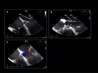
Bicuspid aortic valve Congenital Valvar Aortic Stenosis
Bicuspid aortic valve Congenital Valvar Aortic Stenosis
Congenital Valvar Aortic Stenosis • Patterns of presentation • Presentation in infancy • Almost always severe, rapidly progressive CHF • Untreated mortality : 23% in 1st year • Presentation in childhood • Beyond 1 year of age, heart failure is rare. • Sudden death varies between 1-19%. • Develop progressive ultimately important stenosis • Bacterial endocarditis
Bicuspid Aortic Valve • Natural history • 1. Incidence approximately 1~2% of population • 2. Rarely becomes stenotic or incompetence in early life • 3. Sclerosis begins in the second decade of life. • 4. Aortic stenosis develops in 72% by the fifth & sixth • decades of life. • 5. Endocarditis occurs in 10% of these patients. • 6. Incompetence independent of endocarditis occurs • in 5 - 39% of these patients. • 7. Bicuspid aortic valve have been noted in 25-40% with • supravalvar & 9-20% with subvalvular aortic stenosis.
Bicuspid Aortic Valve • Development • Bicuspid aortic valve, the most common congenital cardiac malformation,is caused by fusion of valve cushions at the onset of valvulogenesis. • At the beginning of valvulogenesis, a population of cells calledneural crest cells migrate away from the neural fold and spreadthroughout the embryo. • These cells seem to play a crucial rolein normal development of cardiac outflow tract &semilunar valves • The basic helix-loop-helix transcriptionfactor dHANDis essential for survival of cellsin neural crest–derived ventricular structures and aorticarch arteries
Congenital Valvar Aortic Stenosis • Techniques of operation • Percutaneous balloon valvotomy • Valvotomy in neonates & critically ill infants • Valvotomy in older infants, children & adults • Aortic valve replacement
Congenital Valvar Aortic Stenosis • Open techniques • Precise commissurotomy • Shaving of thickened leaflets • Excision of obstructive myxomatous nodularities • Mobilization of leaflets • These procedures can be performed with a low surgical risk & 85% freedom from reoperation at 5 years
Bicuspid Valvar Aortic Stenosis • Tricuspidizationwith cusp extension • Criteriafor TCE included an aortic orifice that is equal to or greaterthan normal (normalized for body surface area) after commissurotomyand division of the raphe, adequate mobility of all cusps atthe hinge point, absence of cusp dysplasia involving the bellyof the cusps, commissures that are free of calcification orexuberant fibrosis, and normal location of the coronary ostia. • When these criteria were met, TCE was the procedure of choice.
Bicuspid Aortic Stenosis • Tricuspidizationwith cusp extension The height of coapting pericardial patches is increased toward the neocommissure to compensate for the lack of a true interleaflet triangle and to elevate the hinge point of the leaflets. Each new commissure is constructed by suturing the apposing short edges of each patch together and to the aortic wall, creating an elongated vertical axis of the native commissures
Critical Aortic Stenosis • Optimal management • In fact, if percutaneous balloon valvotomy usually causes rupturealong lines of least resistance, either along underdevelopedcommissures or into leaflet tissue. • Surgical valvotomy allowsdirect inspection of the valve, more fashioning of commissurotomies,and debridement of any excess tissue on the leaflets. • When small aortic annulusand depressed ventricular function are associated, surgicalor therapeutic options other than surgical commissurotomy couldbe considered, including balloon dilation as a bridge to surgery,neonatal Ross operation, Norwood operation, or possibly neonataldouble switch operation.
Bicuspid Aortic Valve • Operative technique
Aortic valve bypass Aortic Stenosis • Aortic valve bypass for high-risk patient with aortic stenosis
Aortic Stenosis • Aortic valve bypass surgery • Ideal candidates for this type of approach could include patientswith ascending aortic calcification, patients who require complexreoperations, and patients with a small annulus • Potential problems include pseudoaneurysm as described in their article,bleeding due to lack of control of the left ventricular (LV)apex, difficulty with the aortic anastomosis in the descendingaorta due to extensive calcification of the descending aorta,kinking of the conduit, and theoretical dislodgement of an LV apical thrombus andnonphysiologic flow from the LV
1. Survival Early deaths Time-related survival 2. Modes of death Acute cardiac failure Sudden death, residual stenosis, incompetence 3. Incremental risk factors for premature death 1) Left-sided cardiac anomalies 2) Preoperative functional class 3) Type of valvar stenosis 4) Young age 4. Functional status 5. EKG changes 6. LV structure and function 7. Residual or restenosis 8. Aortic valve incompetence 9. Bacterial endocarditis 10. Reintervention Results of operation Valvar Aortic Stenosis
Valvar Aortic Stenosis • Indications for operation • 1. Original valvotomy • 1) Neonates and young infants • Treatment on emergency basis • 2) Older infants and children • EKG shows severe hypertrophy • Pressure gradient more than 50mmHg • Symptoms of angina or syncope • 2. Reoperation • Symptoms develop with moderate stenosis
Congenital Aortic Stenosis • Biventricular repair • Contraindications • Small left ventricle < 20ml / BSA, Inlet length < 25mm • Narrow aortic valve ring < 5mm • Small mitral valve orifice < 9mm • Extensive fibroelastosis
Congenital Aortic Stenosis • Norwood vs aortic valvotomy • 1. Mitral valve area less than 4.75 cm 2 /m2 • 2. LV inflow dimension less than 25 mm • 3. Small LV by a ratio between apex-to-base • dimension of LV & that of RV of less than 0.8 • 4. Left ventricular transverse cavity & aortic • annular dimension less than 6 mm
Subvalvar Aortic Stenosis • Introduction • 1. Definition • An obstruction beneath the aortic valve due either to • a short, localized fibrous or fibromuscular ridge or • a long (diffuse) fibrous tunnel. • Subvalvar aortic stenosis may also be a part of other • cardiac anomalies. • 2. History • Chevers : 1st description in 1842 • Brock : Transventricular dilation in 1956 • Spencer : 1st repair using CPB in 1960 • Konno,Rastan : Aortoventriculoplasty in 1975
Subvalvular Aortic Stenosis • Charcteristics • Etiology : unknown but congenital and postnatal • (turbulence phenomenon to abnormal • contractility caused by focal area of • dysplastic myocardium) • Incidence : 10 -20% of AS (0.25 for every 1000 live births) • Anatomy • * Discrete ring of fibrous tissue • * Persistent conus muscle in subaortic area • * Tunnel syndrome( 20% of SubAS)
Subvalvar Aortic Stenosis • Morphology • Aortic valve • Usually normal • Trivial or mild AR in 2/3 due to leaflet thickening, or , • effect of eddy current. • Left ventricle • Usually concentrically hypertrophied • Subendocardial ischemia and fibrosis • Coexisting cardiac anomalies • Isolated in 1/2-2/3 • VSD, IAA, PDA, COA, PS, TOF, ASD, AP window • Other type of discrete subvalvar stenosis • Mitral valve anomalies : accessory tissue or leaflet malposition • Localized muscular obstructions: related to malalignment
Subvalvar Aortic Stenosis • Patterns of type • 1. Localized type • Fibrous or fibromuscular • Localized or circular • Variable degree of septal hypertrophy • 2. Tunnel type • 1/5 of subvalvar aortic stenosis • Circumferential irregular zone of fibrosis • Varying degree of obstruction
Subvalvar Aortic Stenosis Fibromuscular stenosis
Subvalvar Aortic Stenosis Subaortic tunnel stenosis
Subvalvar Aortic Stenosis Supramitral ring
Clinical features & diagnosis Subvalvar Aortic Stenosis • 1. Incidence • 10-20% of AS • 2. Symptoms and signs • 25% requiring operation are asymptomatic. • Systolic murmur, diastolic murmur in 65% • Pulse is slow rising. • 3. Chest X-Ray, EKG • 4. Echocardiography • 5. Cardiac catheterization and cineangiography
Subvalvar Aortic Stenosis • Natural history • 10-30% of congenital LVOT obstruction • Rarely important obstruction in infancy • Evident and progressive with age probably more rapidly than valvar stenosis • Aortic incompetence is a progressive lesion secondary to leaflet thickening.
Subaortic Stenosis • Development & progression • 1. Acquired nature of this lesion • 2. Rarely in neonate and young children • 3. Rheologic theory • Morphologic abnormalities in left ventricular - aorta • junction, such as steeper aortoseptal angle results • in altered septal shear stress and triggers a genetic • predisposition leading to cell proliferation and • structure in LVOT. • 4. Uncertainty about rapidity of progression
Subaortic Stenosis • Development • 1. Subaortic constraint at the entry of the tunnel and the sinus • shape of the letter lead to turbulent flow, resulting in muscle • hypertrophy, or deposition of fibrous material. • 2. Growth of heart without concomitant increase in the size of • VSD, and tunnel • 3. Excessive decrease of LV diameter and the increase in wall • thickness after biventricular repair, causes the diminution of • the VSD orifice and the augmentation of the malalignment. • 4. Other possible causes are kinking of the baffle, shrinkage of • the baffle with time • 5. Chronic flow disturbance caused by a somewhat narrowed • and elongated LVOT
Discrete Fibrous Ring • Histology • In the subaortic region, the progression of discrete fibrous obstruction results in a gross appearance & histology with similarity to vascular lesions by Rodbard. The typical fibrous ring has distinct five layers. • Endothelial layer • Mucopolysaccharide-rich subendothelial layer • Fibroelastic layer • Smooth muscle layer • Central fibrous layer
Subaortic Stenosis Effect of localized stenosis A is normal aorta is depicted with the arrow indicating the direction of flow in the longitudinal view. B, C, and D show the progressive nature of the changes in the aorta.
Subaortic Stenosis LVOT geometry & shear stress ( Aortoseptal angle ) Role of shear stress in the progression of subaortic stenosis
Discrete Subaortic Stenosis • Etiology • Morphologic abnormalities & subsequent rheologic effects, an exuberant • response to local injury, and further exacerbation of the process through • a positive feedback loop
Subaortic Stenosis • Anatomic abnormalities • Increased steepness of aortoseptal angle Malalignment of ventricular septum Prominent ventricular band Protrusion of muscular septum • Increased aorto-mitral separation • Small aortic annulus
Subaortic Stenosis • Extended septoplasty Prior Closure of VSD during Repair of DORV
Subaortic Septal Plane • Anatomy Relationship between the plane of the outlet septum and the plane of the septal crest in the normal heart (A), and atrioventricular septal defect (B), a VSD has been created and a patch applied to augment the diameter of the LVOT.
LVOT & RVOT • Geometry The scheme of the left & right ventricular outflow tract showing the normal anatomy (A), subaortic myectomy (B), a modified Konno procedure (C).