Download
1 / 25
270 likes | 725 Vues
Lectures 21 and 22: The regulation and mechanics of cell division. Today - cell cycle (regulation of cell division) Cell proliferation The eukaryotic cell cycle Measuring the cell cycle Models of the cell cycle: from fungi to frogs The cell cycle is regulated by cyclin-dependent kinases

E N D
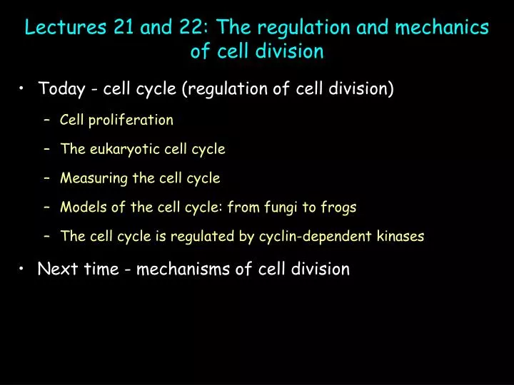
Lectures 21 and 22: The regulation and mechanics of cell division • Today - cell cycle (regulation of cell division) • Cell proliferation • The eukaryotic cell cycle • Measuring the cell cycle • Models of the cell cycle: from fungi to frogs • The cell cycle is regulated by cyclin-dependent kinases • Next time - mechanisms of cell division
A cell cycle is one round of growth and division Cells only come from pre-existing cells cytokinesis mitosis Growth and division must be carefully regulated Unregulated cell growth = cancer
4C 2C 2C 4C The eukaryotic cell cycle is partitioned into four “phases” Division occurs in “M-phase:” “mitosis” and “cytokinesis” (<1 hr) Most cell growth occurs during “G1” (6-20+ hrs; duplicate organelles, double in size) DNA replication occurs during “S-phase” (4-10+ hrs)… “G2” prepares cells for division (1-6+ hrs)… G1+S+G2=“Interphase” Division = “M-phase” A “typical” cell cycle for animal cells is 24-48 hrs long, but varies… 4C (DNA replicated, diploid chr #) 2C (unreplicated DNA, diploid chr #) ECB 18-2
Adapted from MBoC figures 17-5 and 17-6 Number of cells 1 2 DNA content (arbitrary units) Can determine phase of cell cycle from DNA content Where are cells in G1, S, G2 and M on plot? Cells in G1 Which phase has most cells in it? Lasts longest? Cells in G2/M Cells in S ECB 18-2
Transition from one phase to another is triggered We will take a historical perspective to ‘triggers’
Regulating the eukaryotic cell cycle: studies in four model organisms See HWK 618-619 • Marine invertebrates: • Surf clam (Spisula) • Sea urchins and starfish • Frog eggs and embryos: • Rana pipiens (Northern leopard frog) • Xenopus laevis (African clawed frog) • Cultured cells • HeLa (Human cervical carcinoma) • Yeast cell division cycle (“cdc”) mutants: • Saccharomyces cerevisiae “budding” yeast • Schizosaccharamyces pombe “fission” yeast
wee1 cdc25 Phenotype Mutant cdc13 1. Fission yeast “cell division cycle (cdc)” mutants define a master regulator (tigger) of the G2/M transition “Wild-type” fission yeast WT cdc2- (loss of function) “cdc” WEE2 = cdc2D (gain of function) “wee” wee1-(loss of function) wee G2 M cdc2 cdc13-(loss of function) cdc cdc25-(loss of function) cdc Genetic pathway
ECB figure 18-5 Transfer M-phase cytoplasm to interphase oocyte… 2. Frogs: unfertilized eggs contain an M-phase Promoting Factor ECB 18-9 Control expt; Transfer interphase cytoplasm to interphase cell - no effect Nucleus Egg in “M-phase” Oocyte in “interphase” Oocyte “matures” (enters M-phase)… Transfer of cytoplasm from egg to oocyte induces M-phase: “M-phase promoting factor (MPF)” Not restricted to egg cytoplasm - Any M-phase cytoplasm will induce M-phase
M-phase M-phase Interphase Interphase MPF peaks in M-phase MPF activity Time MPF activity cycles during the cell division cycle Peak MPF induces M-phase ECB 18-10
Cyclin A Cyclin B Ribonucleotide reductase (control) M-phase M-phase Interphase Interphase MPF peaks in M-phase MPF activity Cyclin degraded Cyclin synthesis Time 3. Surf clams and sea urchins: the abundance of “cyclin” proteins varies with the cell cycle Continuously label fertilized eggs with 35S-methionine Analyze incorporation into proteins by SDS-PAGE • “Cyclin” abundance varies with cell cycle: • continuously synthesized… • degraded at end of M-phase • Cyclin B mRNA induces M-phase when injected into Xenopus oocytes Peak MPF induces M-phase ECB 18-6
wee1 cdc25 G2 M cdc2 cdc13 Cdc2 gene product is a master regulator of the G2-M transition MPF regulates entry into M-phase Abundance of “cyclins” in clam eggs varies with the cell cycle Three models of the eukaryotic cell cycle • Bringing it all together • Cyclin B mRNA (clam) induces M-phase in frog oocytes • cdc13 encodes a yeast cyclin • MPF consists of frog cdc2 homolog and cyclin B
wee1 P CLB (cdc13) CLB (cdc13) CLB (cdc13) CLB (cdc13) cdc2 cdc2 cdc2 cdc2 P cdc25 P cdc25 (inactive) Cell cycle control: from models to molecules Inhibitory kinase Remove inhibitory phosphate Inactive (weakly active) Active MPF (CDK1) P P CDK1 Inactive Phosphorylate M-phase substrates Histones Lamins MAPs etc Activating kinase Positive feedback ECB 18-11 and 18-12 • “MPF” contains two components: • cdc2gene product =catalytic subunit of protein kinase • cyclin B (CLB = cdc13):regulatory subunit activates kinase • MPF = “Cyclin-dependent kinase (CDK1)” • MPF (CDK1) activity is also regulated by phosphorylation • wee 1 is inhibitory kinase • cdc25 is activating phosphatase “Switching on” CDK1 (MPF) drives cell into M-phase
CLB (cdc13) CLB (cdc13) CLB (cdc13) cdc2 cdc2 cdc2 P P Accumulation of cyclin B APC Inactive APC Active Polyubiquitin MPF triggers its own inactivation“anaphase promoting complex (APC)”; targets cyclin B for degradation Cyclin B accumulation activates MPF MPF activates APC APC inactivates MPF by degrading cyclin B A cytoplasmic oscillator • Metaphase (mid-M) • High cyclin B • MPF (CDK1) active • Prophase (early-M) • Activation of CDK1 by cyclin and cdc25 • Interphase • APC is turned off • Telophase (late-M) • Low cyclin B • MPF inactive • Cyclin B degraded by proteosome • Anaphase
M-phase M-phase Interphase Interphase MPF activity MPF peaks in M-phase Cyclin degraded Cyclin synthesis Time Review: ECB 18-6 Accumulation of cyclin B above threshold activates MPF (CDK1) and promotes entry into M-phase Activation of APC by MPF promotes cyclin destruction, MPF inactivation, and exit from M-phase
Trigger M-phase M-phase cyclin degraded… Active M-phase CDK (MPF) M-phase cyclins (B) M G2 P S-phase CDKs M-phase CDK (CDK1) P G1 S S-phase cyclins degraded… S-phase cyclins Active S-phase CDKs Trigger S-phase At least 6 different CDKs and multiple cyclins in mammals Done M-phase Multiple CDKs regulate progression through the cell cycle G1-CDKs; drive cells through G1 (won’t discuss) S-phase cyclins and CDKs regulate DNA replication Degradation of S-phase cyclins promotes exit from S-phase into G2 ECB 18-13
S-Cdk regulates DNA replication Origin recognition complex - protein scaffolding for assembly of other proteins Cdc6 increases in G1; binds ORC and induces binding of other proteins forming pre-replicative complex Origin is ready to fire ECB 18-14 Active S-Cdk 1- phosphorylates ORC causing origin to fire = replication 2-phosphorylates Cdc6 leading to ubiquitination and degradation Cdc6 not made until next G1 - prevents origin from double firing
Completion of critical cellular processes is monitored at cell cycle “check points” ECB 18-17 • Is DNA undamaged? • Is DNA replicated? • Is cell big enough? • Yes? Enter M phase • Have all chromosomes • attached to spindle? • Yes? Proceed to anaphase • Is the cell big enough? • Is the environment favorable? • Is DNA undamaged? • Yes? Enter S phase Of these, the G1/S checkpoint for damaged DNA is best understood
RNA pol p53 (active) p21 gene Translation Transcription p21 P P The DNA damage checkpoint: p53 induced expression of an S-phase CDK inhibitor p53 (inactive) DNA damage activates p53 Active p53 acts as a transcription factor to turn on genes, including p21 p21 protein inhibits G1/S phase CDKs, blocking entry into S-phase Cell arrests in G1 until damage repaired, or undergoes apoptosis (programmed cell death) DNA ECB 18-15 P21 binds and inactivates S-phase CDK Active S-phase CDK
If checkpoint is activated Exit cell cycle (temporary or permanent) neurons most plant cells Or undergo apoptosis (in a minute)
Zones of division and growth in plant roots Arabidopsis thaliana Only a fraction of cells still actively dividing Zone of differentiation - cells cease growing and terminally differentiate Zone of cell elongation - growth but not division; Cells in G0 Meristem - zone of active cell division cells remain in cell cycle Regulation of each zone is not well understood in plants but involves hormones In animals: mitogens stimulate cell proliferation (block checkpoints) growth factors stimulate cell growth (stimulate biosynthesis, inhibit degradation)
ECB figure 18-19 Tadpole tails are resorbed during metamorphosis ECB figure 18-18 Paws develop from “paddles” Apoptosis: A tale of tadpole tails and mouse pawswhat do they have in common? Both processes involve “programmed cell death (apoptosis)” ECB - “programmed cell death is a commonplace, normal, and benign event. It is the inappropriate proliferation and survival of cells that presents real dangers”
Necrosis (cell death following injury) often results in lysis, spilling the contents into the surrounding space and causing inflamation During apoptosis (“programmed cell death”), cells remain intact and condense Corpses of apoptotic cells are often engulfed by their neighbors or specialized phagocytic cells Apoptosis is visibly distinct from necrosis ECB 18-20
Death protein Inactive Active “Caspases” are proteases; inactive precursors activated by proteolysis Presence of suicide signals and/or withdrawal of needed survival factor activates first caspase in cascade Survival factor Apoptosis is mediated by a “caspase cascade” Caspase (inactive) Initial caspase proteolytically activates downstream caspases …which activate additional caspases, and so on Activated caspases degrade nuclear and cytoplasmic proteins (lamins, cytoskeletal proteins, etc)… Activated endonucleases cut chromosomal DNA… ECB 18-21
Caspase cascade must be carefully regulated Bcl-2 family of proteins are death proteins Form pores in outer mitochondrial membrane releasing cytochrome c (respiratory chain) Cytochrome c binds adaptor and complex activates first procaspase