Download
1 / 22
290 likes | 854 Vues
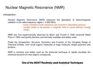
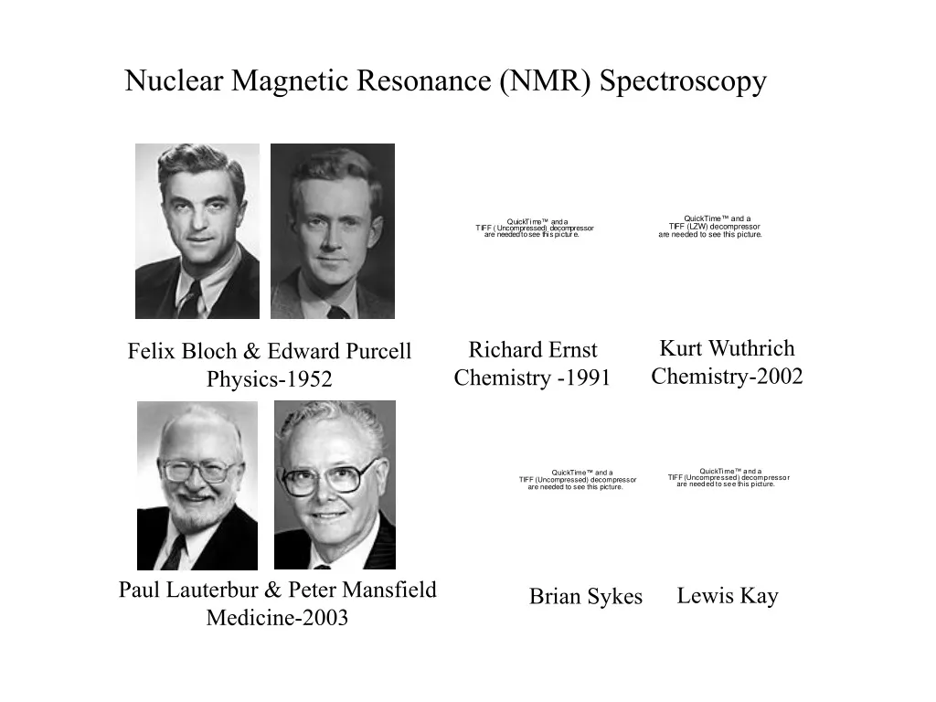
Nuclear Magnetic Resonance (NMR) Spectroscopy. Kurt Wuthrich Chemistry-2002. Richard Ernst Chemistry -1991. Felix Bloch & Edward Purcell Physics-1952. Paul Lauterbur & Peter Mansfield Medicine-2003. Lewis Kay. Brian Sykes. Principles of NMR Protein Spectroscopy. What does NMR tell us ?.

E N D
Nuclear Magnetic Resonance (NMR) Spectroscopy Kurt Wuthrich Chemistry-2002 Richard Ernst Chemistry -1991 Felix Bloch & Edward Purcell Physics-1952 Paul Lauterbur & Peter Mansfield Medicine-2003 Lewis Kay Brian Sykes
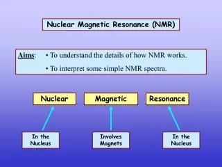
Principles of NMR Protein Spectroscopy What does NMR tell us ? 1) Primary structure characterization 2) Dynamics - psec-sec timescales 3) Equilibrium binding 4) Folding/Unfolding 5) Three dimensional structure Some Advantages 1) Solution based 2) Non-destructive 3) Residue specific information
Crystallography IR UV/Vis NMR Principles of NMR Protein Spectroscopy Wavelength (nm) Radio waves X-rays 10 100 1000 104 105 106 107 108 109 Electron transitions Nuclear spin transitions
Nucleus I # Protons # Neutrons 1H 1/2 1 0 12C 0 6 6 13C 1/2 6 7 14N 1 7 7 15N 1/2 7 8 Principles of NMR Protein Spectroscopy Absorption of energy by nucleus - depends on nuclear spin (I) = sum of unpaired protons + neutrons (spin 1/2) I≠0 - NMR observed - spin will have magnetic moment =I For proteins 1H, 13C, 15N (31P)
Now lets put these in a static magnetic field N S Principles of NMR Protein Spectroscopy Under normal conditions the difference between these is negligible I = 1/2 = Iz = mIh N S Bo N S mI = +1/2 a mI = -1/2 b m=(-I, -I+1 ….I-1, I)
=-Iz z Bo x y In a magnetic field E = -Bo = -gIzBo = -ghmIBo m=(-I, -I+1 ….I-1, I) Principles of NMR Protein Spectroscopy Normal Magnetic Field (Bo)
b DE = ghB0 a 1ghB0 -ghmIB0 - 1ghB0 2 2 Principles of Protein NMR Spectroscopy So how does this give us an “NMR signal” ? N N S N S mI = -1/2 b mI = +1/2 a S E =
b DE = ghB0 a g (*107rad / T sec) 1H 26.75 13C 6.73 15N -2.72 As B0 so does DE ! Principles of Protein NMR Spectroscopy This occurs for all 1H, 13C, 15N in magnetic field B0 B0 = 11.75 T (500 MHz) 14.09 T (600 MHz) 18.79 T (800 MHz)
b DE = ghB0 = hn n = gB0 (Hz) (rad) w = gB0 a Larmor Precession 2p n at 14.09 T g (*107rad / T sec) Nucleus 1H 26.75 600.00 13C 6.73 150.87 15N -2.71 60.82 Principles of Protein NMR Spectroscopy We can use this to select for the nucleus of interest Use frequency n to stimulate transitions !
1H N=106 11.75T 18.79T b 499,968 499,980 DE = ghB0 = hn a 500,020 500,032 Dn = 40 Dn = 64 na-nb = Dn = NghB0 2kT Insensitivity Larger magnet = sensitivity Principles of Protein NMR Spectroscopy NMR is an insensitive method Populations of a, b determined by DE - Boltzman distribution Only measure net difference (Dn)
z z x x y y na-nb = Dn = NghBo 2kT Principles of Protein NMR Spectroscopy Sensitivity Mo=nz + nbz = nz= 1 ghn = 1 N (gh)2Bo 2 4kT Bo Bo - it turns out the observed signal (S) - varies with Bo2
z z o x x Rotating Frame y y Principles of Protein NMR Spectroscopy Net Magnetization Mo Bo Bo
z z x x y y Principles of Protein NMR Spectroscopy How is the NMR Signal Obtained ? -employ a rf pulse at of desired nucleus Mo “rf pulse” Bo B1 B1 2*pw*B1=
z z x x y y Principles of Protein NMR Spectroscopy Mo = /2 Bo B1 B1 2*pw*B1= pw= 1 4*B1 pw = 6 sec B1 = 41 kHz rf off on
z z z x x x y y y Principles of Protein NMR Spectroscopy Mo Bo receiver B1 B1 B1 rf FT on off time
z z z x x x y y y Principles of Protein NMR Spectroscopy Mz(t) = Mo(1-e-t/T1) T1 B1 receiver B1 T2 B1 My(t) = My(0)*e-t/T2 off time
Principles of Protein NMR Spectroscopy T1, T2 (sec) Linewidths 100 10 1 0.1 0.01 0.001 T1 1/2 = 1 T2* T2 0.5 Hz 10 Hz 100 10 1 0.1 0.01 0.001 Correlation Time, c (nsec) MW 20,000 MW 100 Molecular Weight
Principles of Protein NMR Spectroscopy NMR Instrumentation Magnet - Bo 18.79 T vacuum N2(l) He(l) magnet 22.31 T (2006) probe
Field S/N 0.94 T 1 17.62 T 351 22.31 T 563 Ribonuclease (1957) - 40 MHz Lysozyme (1995) - 750 MHz Principles of Protein NMR Spectroscopy Magnet Technology
B1 Bo Principles of Protein NMR Spectroscopy Probe Technology
Principles of Protein NMR Spectroscopy Cold “Cryogenic” Probe S/N ~ 1/{Rs*(Ts+Tpa)+(Rc*(Tc +Tpa)}1/2 Tpa, Tc - lowered 298˚K 20˚K Rs, Ts - near 298˚K 3-4 time more sensitive 500 MHz + cold probe = 1.6x S/N 800 MHz
Principles of Protein NMR Spectroscopy Block Diagram of NMR Spectrometer Probe obs, lk obs, dec, lk Duplexer Transmitter Preamplifier obs, lk Receiver CPU Computer