Download

1 / 16
160 likes | 175 Vues
Simple stain. Rana A.Abo-Hajjras. Simple stain. Simple stain. The simple stain can be used to determine cell shape, size, and arrangement.

E N D
Simple stain Rana A.Abo-Hajjras
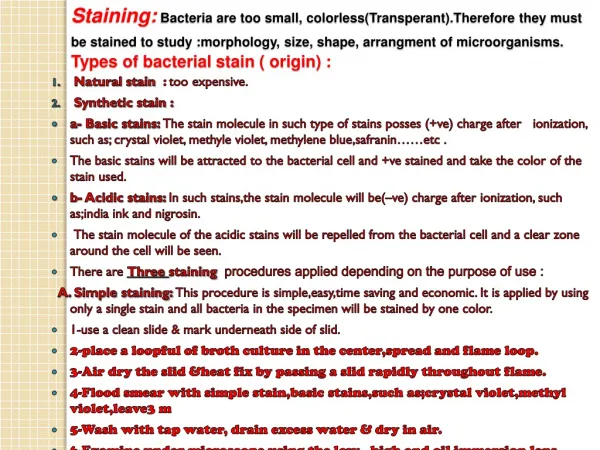
Simple stain The simple stain can be used to determine cell shape, size, and arrangement. True to its name, the simple stain is a very simple staining procedure involving only one stain. You may choose from methylene blue, Gram safranin, and Gram crystal violet.
Simple stain Basic stains, such as methylene blue, Gram safranin, or Gram crystal violet are useful for staining most bacteria. These stains will readily give up a hydroxide ion or accept a hydrogen ion, which leaves the stain positively charged. Since the surface of most bacterial cells is negatively charged, these positively charged stains adhere readily tocell surface
Simple stain A simplestain consists of a solution of a single dye. Some of the most commonly used dyes are methylene blue, basic fuchsin, and crystal violet. Simple stains allow one to distinguish the shape (morphology) of the bacteria. For example, E. coli and Bacillus Subtillus are bacilli or rod-shaped bacteria.
Many bacilli occur singularly, but chains may also be observed. Bacilli very greatly in length and diameter. Staphylococcus aureus and Streptococcus pneumoniae are cocci or spherical bacteria. Cocci may occur singularly, in pairs (as in Streptococcus pneumoniae), or in clusters (as in Staphylococcus aureus). R. rubrum is a spirillum or curved bacterium, Spirilla always occur singularly.
Simple Stain Procedure: • Procedure: • Clean and dry microscope slides thoroughly. • 2. Flame the surface in which the smear is to be spread. • 3. Flame the inoculating loop. • 4. Transfer a loop full of tap water to the flamed slide surface. • 5. Reflame the loop making sure the entire length • of the wire that will enter the tube has been heated to redness.
6. Remove the tube cap with the fingers of the hand holding the loop. 7. Flame the tube mouth. 8. Touch the inoculating loop to the inside of the tube to make sure it is not so hot that it will distort the bacterial cells; then pick up a pinhead size sample of the bacterial growth without digging into the agar. 9. Reflame the tube mouth, replace the can, and put the tube back in the holder.
10. Disperse the bacteria on the loop in the drop of water on the slide and spread the drop over an area the size of a dime. It should be a thin, even smear. 11. Reflame the inoculating loop to redness including the entire length that entered the tube. 12. Allow the smear to dry thoroughly. 13. Heat-fix the smear cautiously by passing the underside of the slide through the burner flame two or three times. Test the temperature of the slide after each pass against the back of the hand. It has been heated sufficiently when it feels hot but can still be held against the skin for several seconds. Overheating will distort the cells.
14. Stain the smear by flooding it with one of the staining solutions and allowing it to remain covered with the stain for the time designated below. Methylene blue - 1 minute Crystal violet - 30 seconds Carbol fuchsin - 20 seconds During the staining the slide may be placed on the rack or held in the fingers. 15. At the end of the designated time rinse off the excess stain with gently running tap water. Rinse thoroughly.
16. Wipe the back of the slide and blot the stained surface with bibulous paper or with a paper towel. 17. Place the stained smear on the microscope stage smear side up and focus the smear using the 10X objective. 18. Choose an area of the smear in which the cells are well spread in a monolayer. Center the area to be studied, apply oil directly to the smear, and focus the smear under oil with the 100X objective. 19. Draw the cells observed.