Download
1 / 63
650 likes | 694 Vues
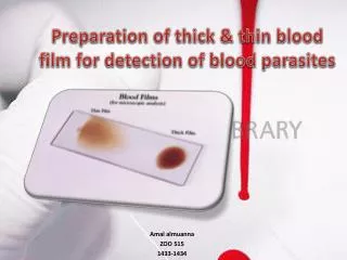
Quality control of Blood film. When blood film needed?. Blood count request: Is it a first time count or repeat count? First time count: Is it a routine screening test or special category? If Routine: Analyzer report for blood count alone Film required if any flags are signaled.

E N D
When blood film needed? • Blood count request: • Is it a first time count or repeat count? • First time count: Is it a routine screening test or special category? • If Routine: Analyzer report for blood count alone • Film required if any flags are signaled
First time count - If Special category, Film required: • Diagnosed blood disease patients • Patients receiving radiotherapy and/or chemotherapy • Renal disease • Neonates • Intensive care unit • If special tests have also been requested for: infectious mononucleosis, haemolyticanaemia, enzymopathy, abnormal haemoglobins • If the clinical details on the request form indicate lymphadenopathy, splenomegaly, jaundice or suggest the possibility of leukaemia or lymphoma • Specific requests by clinician
Repeat count: Film required: • Delta check positive when compared with previous record • Any flag occurs in present count • On each occasion for patients with known blood diseases, for neonates, and when specifically requested by clinicians International Society for Laboratory Hematology
FLAGGING OF AUTOMATED BLOOD COUNTS • "Flagging" refers to a signal that the specimen being analyzed may have a significant abnormality because one or more of the blood count variables are outside specified limits (usually 2SD) or there is a qualitative abnormality that requires a quality control check and/or additional investigation.
Making blood film • Blood film can be prepared from fresh blood without anticoagulant or from EDTA anticoagulanted blood. • blood film should be made on clean glass . • Clear without any dust
Making blood films Three basic steps to make blood film: • Preparation of blood smear. • Fixation of blood smear. • Staining of blood smear.
Optimal blood smear characteristic • minimum 2.5 cm in length terminating at least 1 cm from the end of the slide • Gradual Transition in thickness from thick to thin area ending in a Square or straight edge • No streaks , waves , or troughs
Romanowsky stain • Romanowsky stain→Eosin Y and Azure B) Eosin:Acidic Dye bind to Basic groups (Hb,Granules) → reddish or orange color Azure B: Dye bind to nucleic acid & nucleoproteins →Blue-violet color
Good Peripheral Blood Smear Prepare blood films within 4(3) h of the blood collection in K EDTA. Stain the film within one hour of preparation with a Romanowsky stain, containing fixatives; or fix within one hour with "water-free" (i.e., <3% water) methanol for later staining.
رنگ پس از تهيه از نظر آلودگی قارچی و ميکروبی وهر گونهرسوب و پارتيکل وهمچنين نحوه رنگ گرفتن سلول های خونی بررسی می گردد.رنگ آميزی گسترش های خونی در هر ران کاری موارد فوق بررسی می گردد که بصورت مکتوب ومستند بايد در آزمایشگاه قرار گيرد.کيفيت رنگ آميزی مورد قبول سلول ها مطابق جدول زير می باشد .
Corrected WBC Nrbc/100 wbc If 10 (5) or more Nrbc are observed , corrected WBC must be calculated
DIFFERENCES BETWEEN CAPILLARY & VENOUS BLOOD • The differences may be exaggerated by cold with resulting slow capillary blood flow. • The PCV, RBC, and Hb of capillary blood are slightly greater than in venous blood. • The WBC & neutrophil counts are higher by about 8%; • The monocyte count is higher by about 12%, and in some cases by as much as 100%, especially in children. • Conversely, the platelet count appears to be higher in venous than in capillary blood; this is on average by about 9% and in some cases by as much as 32%. • This may be the result of adhesion of platelets to the site of the skin puncture.
Hypochromia (correlate with MCHC) • 1+ :area of central pallor is ½ of cell diameter • 2+ : area of central pallor is 2/3 of cell diameter • 3+ : area of central pallor is ¾ of cell diameter • 4+ : thin rim of hemoglobin
انجام گروه خونی به دو روش : • Cell Type • Back Type • توسط 2 نفر • انجام تست Du جهت گروه های خونی Rh (-) :
stain precipitate smudge cells leukemia