Download
1 / 38
410 likes | 809 Vues
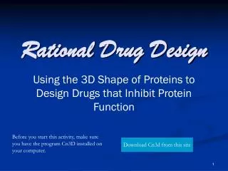
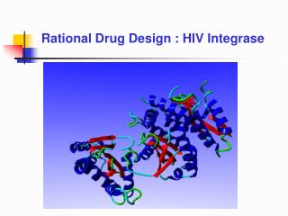
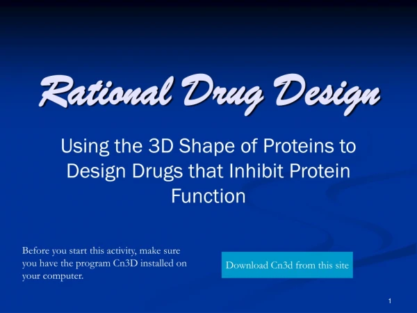
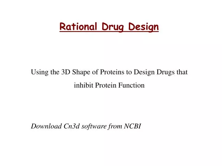
Rational Drug Design. Using the 3D Shape of Proteins to Design Drugs that inhibit Protein Function. Download Cn3d software from NCBI. Why is protein structure important?. Each protein molecule has a characteristic 3D shape that results from coiling and folding of the polymer chain.

E N D
Rational Drug Design Using the 3D Shape of Proteins to Design Drugs that inhibit Protein Function Download Cn3d software from NCBI
Why is protein structure important? • Each protein molecule has a characteristic 3D shape that results from coiling and folding of the polymer chain. • The function of a protein depends upon the shape of the molecule.
Protein chains Each protein has a specific sequence of amino acids that are linked together, forming a polypeptide
The protein chain folds • Interactions between amino acids in the chain form: • Alpha helices • Beta sheets • Random coils Together usually form the binding and active sites of proteins
And folds again.. • After folding, amino acids that were distant can become close • Now the protein chain has a 3D shape that is required for it to function correctly
The final protein…3D structure • The final protein may be made up of more than one polypeptide chain. • The polypeptide chains may be the same type or different types.
Examples of Protein Function Hormones Insulin binds to receptors on cell membranes signalling cells to take up glucose from the blood Protein ChannelsRegulate movement of substances across the plasma membrane. E.g. The CFTR (cystic fibrosis transmembrane conductance regulator) protein pumps ions across membranes Transport Haemoglobin (far right) in red blood cells transports oxygen to cells around the body
Catalase - enzyme action • Hydrogen peroxide, a natural product of metabolism in your cells, is highly toxic in high concentrations and must be removed quickly! Reactants Products oxygen Add ferric ions (Fe 3+) Rate increases 30 000-fold Add Catalase Rate increases 100 000 000-fold water Hydrogen peroxide Location of active site where Hydrogen peroxide binds
Ripping things apart Joining things together How enzymes do it? • Enzyme proteins have specific sites where all the action happens. We call this the active site. Molecules that need to be ripped apart or put together enter the active site. • Each protein has a specific shape so it will only perform a specific job.
Influenza Pandemics The Spanish Fluin 1918, killed approximately 50 million people. It was caused by the H1N1 strain of influenza A. The Asian Flu in 1957 was theH2N2 influenza A strain. Worldwide it is estimated that at least one million people died from this virus. The Hong Kong Flu in 1968 evolved into H3N2. 750,000 people died of the virus worldwide
Designing a Flu Drug Step 1: looking for protein targets Influenza viruses are named according to the proteins sticking out of their virus coat. (H) There are two types of protein = N and H. N and H have special shapes to perform specific jobs for the virus. (N)
N cuts the links between the viruses and the cell surface so virus particles are free to go and infect more cells. H attaches to cell surface proteins so virus can enter cell Virus Proteins on cell surface Virus genes are released into the cell. The lung cell is ‘tricked’ into using these genes to make new virus particles. Human Lung Cell
Blocking the active site Neuraminidase in Cn3D RELENZA This link will open a Cn3D file of Neuraminidase with the drug relenza blocking its active site Australian team did it
Some facts… • Calcium, sodium and potassium ions control essential functions inside cells: calcium, for example, helps regulate the contraction of muscle cells. • Ion channels control the entry and exit of ions into and out of cells. • Some conotoxins act as analgesics, interacting with ion channel receptors in nerves so the ion channel cannot open. Blocking ion channels stops ions from entering a neighbouring nerve fibre. No electrical impulse is set off so the ‘pain’ message is switched off!
Sodium ion Calcium ion Acetylcholine The nerve impulse 3.Influx of Calcium causes acetylcholine to be released into synaptic junction. Synaptic Junction Na+ Ca2+ + + - - 2. Sodium ions accumulate causing Calcium ion channels to open. - - + + 4. Acetylcholine binds with receptor proteins changing the shape of the ion channel. 5. This opens the sodium ion channel to let in sodium. 6. Sodium ions set off an electrical impulse along the next nerve cell. 7. The pain message is working. 1. Electrical impulse generated along axon – sodium ions (red) rush in and Potassium ions (green) rush out To block pain we can try to target the ion channels.
Na+ ion channel You will explore this part of the ion channel. This is the section that binds acetylcholine &/or drug molecules causing the ion channel to change its shape. Outside neuronal cell Cell membrane (Phospholipid bylayer) Inside neuronal cell Some conotoxins block acetylcholine (nACh) receptors that stud the surface of neurons.
Chemoinformatics QSAR • Quantitative Structure Activity Relationship (QSAR) is a set of methods that tries to find a mathematical relationship between a set of descriptors of molecules and their activity. • The descriptors can be experimentally or computationally derived. Using regression analysis, one can extract a mathematical relationship between chemical descriptors and activity.
Descriptors in QSAR study • Constitutional Descriptors • Topological Descriptors • Geometrical Descriptors • Electrostatic Descriptors • Quantum Chemical Descriptors • Thermodynamic Descriptors • Reactivity Descriptors
Constitutional Descriptors • Total number of atoms in the molecule • Absolute and relative numbers of atoms of certain chemical identity (C, H, O, N etc.) in the molecule • Absolute and relative numbers of certain chemical groups and functionalities in the molecule • Absolute and relative numbers of single, double, triple, aromatic or other bonds in the molecule • Total and relative number of 6 membered aromatic rings • Molecular weight and average atomic weight
Topological Descriptors • Molecular connectivity index • Valence connectivity indices • Shape indices • Flexibility index • Structural information content index • Bonding information content index
Geometrical Descriptors • Molecular surface area • Solvent-accessible molecular surface area • Molecular volume • Solvent-excluded molecular volume • Principal moments of inertia of a molecule • Shadow areas of a molecule • Relative shadow areas of a molecule
Electrostatic Descriptors • Atomic partial charges • Minimum (most negative) and maximum (most positive) atomic partial charges • Polarity parameters • Dipole moment • Average ionization energy • Minimum and maximum electrostatic potential at the molecular surface • Local polarity of molecule • Total variance of the surface electrostatic potential • Electrostatic balance parameter
Quantum Chemical Descriptors • Total energy of the molecule • Standard heat of formation • Electron-electron repulsion energy for a given atomic species • Nuclear-electron attraction energy for a given atomic species • Nuclear repulsion energy between two given atoms • Total intramolecular electrostatic interaction energy • Electron kinetic energy density
Problems with QSAR descriptors • Ideally, chemicals with similar descriptors should show similar activity but similar in one context may not mean similar in another. • Difficult to represent interactions between descriptors • With many different types of descriptors, how do you compare them (e.g. what does it mean if the shapes are highly similar, but the charge distributions are very different?)
PubChem Compound http://pubchem.ncbi.nlm.nih.gov/
Sanjeevini Software Sanjeevini software has been developed to provide a computational pathway for automating lead design. The user can upload a bimolecular (protein) target and a candidate drug. The software identifies the potential active sites, docks and scores the candidate drug and returns four structures of the candidate drug bound to protein target together with binding free energies. The Ligand and Protein should be provided in pdb format for all the modules.
Modules of Sanjeevini software • NRDBSM database • Aimed specifically at virtual high throughput screening of small molecules and their further optimization into successful lead-like candidates. • It has been developed giving special consideration to physico-chemical properties and Lipinski's rule of five, which determines the solubility, permeability and transport characteristics across membranes. • Some of these are molecular weight, number of hydrogen bond donors and acceptors, and molar refractivity. Fixed precincts for these properties have been employed as filters to assemble the database. • Currently holds close to 17,000 molecules with simple structures, low molecular weight, less number of rings and rotatable bonds, low hydrophobicity such that after screening, optimization and consequent increase in molecular complexity, they would show a drift towards 'drug-like' property space.
Weiner Index • The ability of protein to bind to its substrates and to inhibitors in a highly specific manner is an important feature of many biological processes. • For example, determination of binding free energy of the target (protein) with a ligand (any small molecule that binds protein) is a very important step to design new drugs, determination of toxicity of certain chemical materials on living organisms, and many other pharmaceutical and biochemical application developments. • The most crucial bottleneck to finding binding free energy of the target with a ligand is the computational time. For example, it requires more than a minute to find the binding free energy of a target (protein) with only a single ligand in rigid docking method. • For flexible (non-rigid) docking method, it may require several minutes for a single candidate only. There are millions of such ligands which need to be tested to determine the good candidate(s) for a particular target protein.
Lipinski Rule of Five • Lipinski rule of 5 helps in distinguishing between drug like and non drug like molecules. • It predicts high probability of success or failure due to drug likeness for molecules complying with 2 or more of the following rules - • a) Molecular mass less than 500 Dalton • b) High lipophilicity • c) Less than 5 hydrogen bond donors • d) Less than 10 hydrogen bond acceptors • e) Molar refractivity should be between 40-130 • - Molar refractivity is a measure of the total polarizability of a substance and is dependent on the temperature and the pressure. • These filters help in early preclinical development and could help avoid costly late-stage preclinical and clinical failures
Charge Derivation • Partial atomic charge is very crucial for computing physical, chemical and biological properties, and reactivity of molecules. • Through the information of the atomic charge in a given species it is possible to predict the stability, solvation energetics of various molecules, and course of a particular reaction, determine its interaction with biological molecules and so on. • The usefulness, notwithstanding, there is no direct method to determine the partial atomic charges from experiment. During the last few decades various methods have been developed to determine the partial atomic charges, but all these methods have their limitations. • Two methods can be used for the partial charge derivation: TPACM4 or AM1BCC
TPACM4 • Transferable Partial Atomic Charge Model – up to 4 bonds is used for deriving the partial atomic charges of small molecules for use in protein/DNA-ligand docking and scoring. • The main idea of TPACM4 is based on a look up table of template fragments consisting of 4-bond paths around the atom being charged. • This method overcomes the limitations of time complexity of assigning the partial atomic charges of a given molecule. The low value of average error against an experimentally observable physico-chemical properties indicates the reliability comparable to the RESP/AM1BCC results.
Active site finder: The active site finder finds all the possible cavity points in a protein biomolecule along with the residues which are lining the cavity. The user can identify the cavity of interest on the basis of the amino acid lining the cavity and dock the candidate molecule in the cavity.
Software developed 1. SVMProt: Protein function prediction software http://jing.cz3.nus.edu.sg/cgi-bin/svmprot.cgi 2. INVDOCK: Drug target prediction software 3. MoViES: Molecular vibrations evaluation server http://ang.cz3.nus.edu.sg/cgi-bin/prog/norm.pl
Bioinformatics database developed 1. Therapeutic target database http://xin.cz3.nus.edu.sg/group/cjttd/ttd.asp 2. Drug adverse reaction target database http://xin.cz3.nus.edu.sg/group/drt/dart.asp 3. Drug ADME associated protein database http://xin.cz3.nus.edu.sg/group/admeap/admeap.asp 4. Kinetic data of biomolecular interactions database http://xin.cz3.nus.edu.sg/group/kdbi.asp 5. Computed ligand binding energy database http://xin.cz3.nus.edu.sg/group/CLiBE/CLiBE.asp