Download
1 / 48
480 likes | 642 Vues
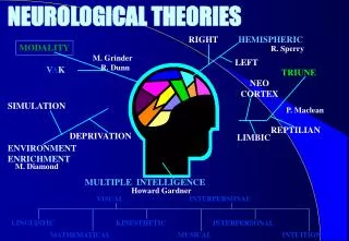
Hemispheric Specialisation. Left side V’s Right side. Left – work Right –play. Hemispheric Specialisation. Left – Verbal Analytical (movement and sensation right side of body, including sight from right Visual field).

E N D
Hemispheric Specialisation Left side V’s Right side
Hemispheric Specialisation • Left – Verbal Analytical (movement and sensation right side of body, including sight from right Visual field) • Right – Non Verbal, Visio Spatial, Music, (movement and sensation left side of body, including sight from left Visual field)
The cross over • Left controls Right • Right controls Left • No one knows why so don’t ask! • This diagram is from the back • Note sexy yellow budgie smugglers
The Reticular Activating System Wake up…. Go to sleep….wake up…..go to sleep Look over there!
BRAIN STEM • Spinal cord is a continuation of the brain stem (also called “hindbrain”) • Brain stem is a structure that looks like a stem on which the brain sits. • Three anatomical structures comprise the brain stem: • Medulla • Cerebellum • Pons Copy Figure 4.31 on page 204
RETICULAR FORMATION • Runs up through centre of brain stem, upward through midbrain to the forebrain (area under the cerebral cortex) is a structure known as the reticular formation. • Through a microscope it looks like white netting or lacing (reticular means “like a network). • Neuroscientists Moruzzi and Magoun (1949) electrically stimulated reticular formation of cats and they woke suddenly and remained alert. When severed connections between reticular formation and remainder of brain the cats fell into a prolonged coma until they died. • Moruzzi and Magoun (1949) thus proposed the function of the reticular formation was to control sleeping and waking.
RETICULAR FORMATION • From the influence of these findings, the reticular formation gradually came to be known as the “reticular activating system”. • It is now recognised there is more to this structure than regulating sleep and wakefulness and that other parts of the brain are also involved in regulating wakefulness. • Exactly what the reticular formation does and how it does it is still unclear.
RETICULAR ACTIVATING SYSTEM • It is a network of neurons that extends in many directions from the reticular formation to different parts of the brain and spinal cord (like a bicycle wheel hub). • Ascending tracts (upward nerve pathways) extend to the cerebral cortex and descending tracts (downward nerve pathways) extend to the spinal cord.
The Reticular Activating System • Major functions are to regulate cortical arousal (“alertness”) either increasing or decreasing arousal in response to feedback from upper and lower brain areas. • Helps regulate sleeping and waking – anaesthetics operate by dampening RAS neural activity • Less active RAS we go to sleep. • Damage to the RAS leads to permanent coma or a chronic vegetative state. • Plays a major role in selective attention – helps decide which stimuli will enter awareness • Steady stream of impulses from RAS keep cerebral cortex active and alert. • Filters incoming sensory information – helps us ignore useless information (weak or familiar sensory information) • Highlights neural information that is of importance, directing attention to potentially significant events. E.g. Country driver sees a kangaroo in middle of road.
RETICULAR ACTIVATING SYSTEM • When event occurs that demands our attention, RAS bombards cortex with stimulation to arouse specific cortical areas. • Information from 2 simultaneous sensory sources (e.g. Sounds and images) the RAS calls our attention to both and assists cerebral cortex to focus on the more relevant information. • Brain scanning shows activation when shifting attention between stimuli • Helps regulate cardiovascular system in response to external stimuli – eg. Sympathetic arousal • Ascending RAS tracts connect to central thalamus areas and appears to influence arousal and attention through the thalamus.
The Thalamus What should I pay attention to? What will enter my conscious awareness?
The Thalamus • Located in middle of the brain, right on top of the brain stem. • About 3 cm in length, consisting of 2 ovoids (look like 2 little footballs) • Each ovoid in a different hemisphere. • Due to location, sometimes referred to as the “gateway” from lower part of brain to the cortex in the upper part. • Sensory relay station – filters information from senses and transmits info to cerebral cortex. • Receives input from all major senses, except smell, which has a direct route to the cortex, bypassing the thalamus. All other sensory info must pass through thalamus to reach the cortex. • Integrates information from the senses – puts the voice with the face and the handshake • Damage can cause sensory problems – blindness, deafness etc. (except smell) or cause the cortex to misinterpret or not receive sensory info. • Some form of analysis or processing of sensory information is likely to occur in the thalamus (based on evidence from case studies of brain damaged patients or those with an abnormal thalamus).
The Thalamus • Closely connected to the RAS and thus also very involved in arousal &attention – does the shifting and filtering • Streams or bursts of information are channelled from RAS through thalamus • Also controls sleeping and waking – arousal through connection to reticular formation and nerve pathways of the RAS. Damage resulting in lower arousal, lethargy or coma. • Info from cerebral cortex also travels through thalamus to lower brain structures, spinal cord and out to peripheral nervous system. E.g. Thalamus has neurons that relay messages between motor cortex and movement control centres in brain stem (such as cerebellum). • Involved in consciousness – sleep-wake, attention, blocks info to the brain while sleeping • Also involved in emotional responses (interpretations) to stimuli • Also linked to disorders like Narcolepsy , Depression and Schizophrenia (misinterpreting or not noticing some stimuli)
The Thalamus • Plays a role in attention by actively filtering vast amounts of incoming info that needs to be attended to, highlighting and giving weight to some inputs and less to others. LaBerge and Buchsbaum (1990) one condition participants attend to a single letter’s presence or absence, in the other condition participants had to “look out” for same letter embedded among other letters. Greater PET activation of a specific area of thalamus was shown in 2nd condition, even when accounting for stimulus complexity.
The spinal cord Neural information super Freeway
The Spinal cord – linking the CNS to the PNS • Spinal cord is cablelike column of nerve fibres extending from base of brain to lower back, encased in a series of bones called vertebrae that extend further than actual cord. • Major function of spinal cord is to receive sensory info from body (via PNS) and transmit them to the brain, then to receive info. from brain and relay it to the body (via PNS) to control muscles, blands and internal organs. • Spinal cord is the super highway for all information / messages coming to (afferent - sensory) and leaving (efferent - motor) the brain. • There are separate pathways for information / messages coming to and leaving the brain – these are called tracts • Damage to spinal cord causes brain to lose both sensory input from the body as well as body control. • Severity of feeling loss and paralysis depends on location of spinal cord damage (higher up spine the greater number of nerve connections between brain and body are severed).
The Spinal Cord • The spine contains two types of matter • White Matter = Myelinated axons solely that run length of spinal cord uninterrupted and are bundled together (part of neuron that sends information away from cell body or soma) Myelin is a protective coating that helps speed up transmission of information • Interconnected axons in CNS are referred to as tracts, circuits or pathways. • Spinal cord has ascending tracts for somatosensory information and descending tracts for motor information. • Grey matter contains neuron bodies (cell bodies, axons and dendrites), mainly located near the centre of the spinal cord.
The Spinal Cord • Adult spinal cord is only about 43 to 45 cm long but vertebrae extend for about 60 cm.
The Spinal cord - levels • Four Sections of Cord – Cheese (cervical) Tastes (thoracic) Like (lumbar) Shit (sacral) • These sections of the spinal cord are named by the groups of nerves that enter and exit the spinal cord at each section • Cervical – 8 pairs of nerves enter and exit • Thoracic – 12 pairs of nerves enter and exit • Lumbar – 5 pairs enter and exit • Sacral – 5 pairs enter and exit • Nerves at each level send / receive information to / from different parts of the body (see handout) • The spinal cord only goes to the base of the thoracic vertebrae. Nerves radiate laterally from the spinal cord above this, and continue vertically inside the vertebrae below the thoracic – this bundle of nerves is known as the caudaequinaor horses tail
The Spinal Cord • Cervical nerves – each level contributes to different motor functions of neck, shoulders and arms. • Thoracic nerves – Radiate to muscles in chest (pectoral muscles). Also links to visceral muscles which are connected to large internal organs in chest cavity (e.g. lungs). Thus involved in actions such as breathing and coughing. • Lumbar nerves – Leg muscles • Sacral nerves – Bowel, bladder and sexual function • Spinal cord only extends to last bone of thoracic vertebrae. Nerves below thoracic section form a bundle running vertically inside vertebrae. This large section of nerves is known as the caudaequina (“horse tail”).
The Spinal Reflex • 1730 English scientist Stephen Hales – demonstrated spinal cord responsible for limb reflex movements. • Decapitated a frog (thus no brain input) and pinched a leg, the frog’s leg pulled away. • Hales concluded sensory receptors in leg had transmitted message to spinal cord and in turn message looped to motor neurons to stimulate certain muscles, causing contraction and pulling away from stimulus. • Pain not registered as pain registers in the brain. • Interneuron cells in spinal cord connect between sensory neurons and motor neurons to create the loop. • The same reflex response occurs in humans to protect from harm and enhance survival. E.g. Prick finger on rose thorn.
Neurons –building blocks of the Nervous System • A neuron is a cell that sends and receives information in the form of neural impulses (neural impulses are tiny pulses of electro chemical energy) • Sensory and motor neurons do not share the same tracts to and from the brain, they follow separate but adjacent paths • 31 afferent and 31 efferent tracts in spinal cord (separate train tunnels) • Sensory info is transmitted from senesory receptor cells in PNS via sensory neurons to the CNS. • Sensory information is “afferent” information (afferent = coming towards) i.e. Towards CNS e.g. Hearing a kettle whistle, seeing a bird fly, feeling cold of an ice cube. • Sensory (feeling) neurons – afferent (coming towards) • Motor (Moving) neurons – efferent (leading out) from CNS • Interneurons – connecting neurons that relay messages from sensory neurons to other interneurons or moto neurons within the CNS (enable the spinal reflex, sensory communicate with motor)
Transmission of neural information • Motor information from the brain comes from motor cortex at rear of frontal lobes (except for spinal reflex) where motor neurons carry messages of movement down the spinal cord to the skeletal muscles and glands where they connect to effector cells. • Effector cells control action of muscles by causing them to contract, thus causing movement. They are located in glands and cause the secretion (release) of hormones to occur when they are activated by messages from motor neurons.
Neurons • There are many types of neurons – about 200 different in the CNS alone. • Can be categorised into 3 types based on their primary function: sensory, interneurons, motor neurons. • Sensory neurons – afferent neurons. Carry messages from sensory organs through nerves in PNS up tracts in spinal cord to the brain. Receive info. from both external and internal environment. E.g. Environment through senses and internally from muscles, organs and glands. Sensory information known as afferent information. • Nerve tracts in the spinal cord that carry this information carry ONLY somatosensory information. • Sensory neurons are quite different in structure to motor neurons and DO NOT share the same tracts to and from the brain. • There are 31 afferent tracts and 31 efferent tracts in the spinal cord. • Draw a sensory neuron and a motor neuron (see page 210) • Some sensory info. transmitted to brain from spinal cord. • Some sensory info. directly transmitted to brain from cranial nerves in face (e.g. From forehead or cheek). • Sensory neurons usually only respond to a particular type of stimulation. E.g. Neurons in nose to odours detected by chemical energy but not light (electromagnetic energy) or sound (mechanical energy).
Transmission of sensory info from the Peripheral Nervous System • Afferent means “coming towards” – in this case coming towards the Central Nervous System. • Information such as change in skin temperature when holding an ice cube, sound of a kettle whistling, smell of toast cooking are all examples of sensory information.
Interneurons • Interneurons – connecting neurons located near the base of the spinal cord. Make connections between sensory and motor neurons which rarely ever connect directly or to other interneurons. Exist only in CNS where they are in largest number of any type of neuron. Can have long axon and short dendrites or short axon and long dendrites. Draw an interneuron (see page 210)
Transmission of motor information from the brain • After the brain receives sensory stimuli information there is often a response that requires movement. • The motor cortex is the “launching pad” for most bodily movements, whether voluntary or involuntary (except movement initiated by the spinal reflex). • Motor neurons are the neurons that carry messages of movement. They leave the motor cortex (at the rear of the frontal lobes), travel down the spinal cord to the skeletal muscles and glands where they connect to effector cells. • The effector cells control the action of muscles by causing them to contract, thus causing movement. Effector cells located in glands cause the secretion (release) of hormones to occur when they are activated by messages from motor neurons.
Motor Neurons • Motor neurons – efferent neurons. Carry messages away from CNS towards muscles, organs and glands, thus enabling body movements, internal organs to be activated and glandular secretions to occur. • Motor messages are known as efferent messages. • Efferent means “leading out from” – in this case, leading out from the central nervous system. • Physically turning taps when shower is sensed as too hot are messages transmitted from the motor cortex, down the spinal cord, out to effector cells located in the muscles of arms, hands and fingers. • Direction of neural impulses and what happens at destination different to sensory neurons.
Cerebral Cortex • There is ongoing interaction in the cerebral cortex between sensory information coming to the brain and motor information going from the brain because a lot of the sensory information requires action to be taken in the form of movement. • The close proximity of the somatosensory and motor cortices has been suggested as an adaptive evolutionary feature of the brain.
A Motor Neuron Terminal Button Axon terminals synapse Myelin sheath Axon
STRUCTURE OF A NEURON • The soma or cell body is the structure that determines whether the neuron will be activated and thus transmit (send) messages to other neurons. • A dendrite (from the Greek work meaning 'tree') is a short, thin, widely branching nerve fibre that is specialised to detect and receive neural information. • An axon is a single, tube like, fluid-filled extension that transmits messages from the soma to other cells in the body including other neurons, muscles, organs and glands. Encased in a white fatty substance that helps speed un transmission called myelin sheath. • At the end of each axon are branches called axon terminals. Each axon terminal has a small knob-like swelling at the tip of it called a terminal button / synaptic knob • The synaptic knob is a small structure like a sac that stores chemicals called neurotransmitters which assist in the transmission neural information from one neuron to another
The Neural Impulse • Neurons are capable of only transmitting information in a specific form, as neural impulses. • A neural impulse (or “action potential”) is a combination of electrical and chemical energy (electrochemical energy) that contains the neural information that travels along the axon. • A neural impulse travels only the length of a neuron and not continuously along the axon. When impulse is triggered, one section of axon opens up that triggers the next section to open up and so on passing the impulse along the axon • Between membrane casing of neuron and the neuron is a small amount of fluid containing particles called ions.
The Neural Impulse • The ions are either positively charged or negatively charged with different quantities of each in the fluid. • The difference in the charges between the neuron and the surrounding fluid results in an electrical charge across the cell membrane. • Neuron in resting state there is a very small difference in electrical charge inside and outside the neuron. The difference is known as the resting potential. • Each neuron requires a minimum level of stimulation to be activated from its resting potential so a neural impulse begins. This minimum level to activate a neural impulse is the neuron’s threshold.
The Neural Impulse • Resting potential changes when a neuron receives messages from other neurons. If the electrical charge reaches the threshold, a neural impulse is activated and begins its movement down the axon, in sections. • At the end of each axon section is an ion channel. Ion channels are like gates that open and close to allow ions to flow along the axon. • The neural impulse can only move in one direction. • Once triggered, a neural impulse is self-sustaining – continuing to the end of the axon without further stimulation. • No matter how much higher than the threshold, the neural impulse will occur. Magnitude (intensity) is not important, there is either a neural impulse or not (i.e. It either fires or doesn’t fire). There is no such thing as a “partial impulse”. This rule is called the “all or nothing principle”. • Neural impulse speed down an axon varies. Fastest is 430 km/h and slowest at 3.5 km/h. Speed depends on diameter (width) of the axon and whether it has a myelin sheath. Larger diameter = faster, myelinated axons = faster.
Synaptic transmission • The gap between neurons is called the synapse. • When the neural impulse reaches the end of each axon, the terminal buttons releases chemicals called neurotransmitters • A neurotransmitter is a chemical substance that is manufactured by the neuron. It contains ions that travel across the synapse to the receptors on the dendrites of the receiving neuron • Sometime the neurotransmitter triggers or activates a neural impulse on the connecting neuron. At other times, the neurotransmitter inhibits or prevents the connecting neuron from firing • When the neurotransmitter has done its job it is either taken back by the terminal buttons or disposed of
100 billion neurons 10,000 connections for each neuron
The Parts of a Neuron • Dendrites • Cell body (soma) • Axon • AxonTerminals • Terminal Buttons • Synapse • DCATBS • Don’t Confuse All This Bull Shit! • Do CAT’sBite Snakes?
Parts of a neuron story? • Denny Dendrite was a prisoner who lived in a cell with a dead body. He was in jail for murdering his mother with an axe on her way to the train terminal. He also likes to collect buttons?
Stuff to do • Hemispheric dominance quiz • Handout on lobes • Neuron structure handout • Learning activities – 4.9, 4.10, 4.12, 4.13