Download
1 / 12
140 likes | 353 Vues
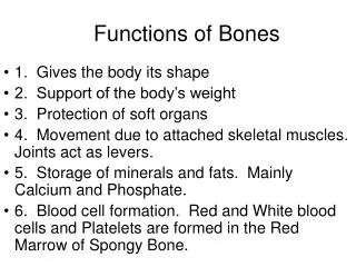
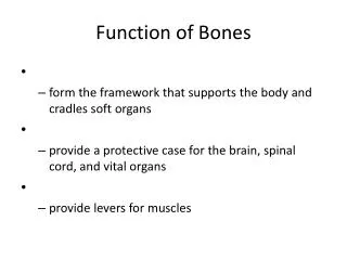
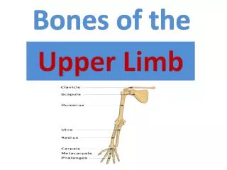
Anatomy of Bones. Head 1.Frontal 2 occipital 3 parietal 4 Temporal Neck and spine Cervical=C1-C7 Thoracic=T1-T12 Lumbar= L1-L5 Sacrum Coccyx. Upper extremity Clavicle Scapula humerus Ulna Radius Carpals Metacarpals phalanges Torso Sternum Xiphoid process Ribs (12). Bones.

E N D
Head 1.Frontal 2 occipital 3 parietal 4 Temporal Neck and spine Cervical=C1-C7 Thoracic=T1-T12 Lumbar= L1-L5 Sacrum Coccyx Upper extremity Clavicle Scapula humerus Ulna Radius Carpals Metacarpals phalanges Torso Sternum Xiphoid process Ribs (12) Bones
Bones continued • Lower extremity • Pelvis • Femur • Patella • Tibia • Fibula • Tarsals • Calcaneous • Talus • Metatarsals • Phalanges
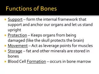
Anatomy • Types of Bones • Long-curved and long • Femur, humerus, tibia, fibula, radius, ulna, phalanges, metatarsal/metacarpal • Most commonly injured • Short- cubed shaped, they are spongy bone except at the surface, • Wrist- carpals • Foot- tarsals • Flat- thin and composed of two nearly parallel plates of compact bone • Ex. Cranium, sternum, ribs, scapula • Irregular- compact - vertebrae • Sutural or Wormian- small bones located between the joints of certain cranial bones. Soft spot on your head
Types of bones cont. • Sesamoid- small bones wrapped in your tendons • 1st metatarsal • Spongy- short, carpals, tarsals • Compact- vertebrae
Anatomy of bone Structure • Diaphysis- main shaft of the long bone- hollow and covered by compact bone or periosteum • Epiphysis-end of long bones provide for muscle attachments • Metaphysis- the wider part at the end of the long bone- adjacent to the epiphyseal plate • Articular cartilage-hyaline cartilage- joint surfaces • Periosteum- covering of long bone. Interlacing with the periosteum are fibers from the muscle/tendons • Medullary-hollow tube in the long bone contains a yellow fatty marrow • Endosteum- lining the medullary cavity
Type of joints • Gliding- where bones glide over one another. • Ex. Vertebrae, tarsals, carpals • Hinge- one surface is round that fits into another concave surface. • Ex. Elbow, knee: motion flex/ext • Ball and Socket-movement in many planes/axis, such as ext/flex, int/ext rotation, abd/adduction, circumduction • Ex. Hip and shoulder • Condyloid-movement similar to ball/socket but in two planes; forward/backward, flex/extension, abd/adduction • Ex. Wrist, metacarpal phalangeal joint • Saddle- flex/extension, add/abduction circumduction • Ex. thumb • Pivot- rotation-movement in one plane about one axis. • Ex. Radio-ulnar joint
Anterior/Posterior Medial/Lateral Superior/Inferior Supine/Prone ______________ 1 flexion 2 extension 3 abduction 4 adduction 5 internal rotation 6 external rotation 7 pronation 8 Supination 9 Dorsi flexion 10 plantar flexion 11 Circumduction 12 inversion 13 Eversion 14 horizontal flexion/extension 15 protraction/retraction Anatomical Movements
ROM is predetermined by: • 1.Flexibility of muscles • 2.flexibility of tendons • 3.Flexibility of ligaments • 4. By size • 5.Factors of use • 6.Injury • 7.Age • 8.Angle of the joint • 9.Disease
Rectus Parallel to the midline Ex. Rectus abdominis, rectus femoris Transverse Perpendicular to the midline Ex. Transverse abdominis Oblique Diagonal to the midline Ex. External Oblique or Internal oblique Maximus Large Ex. Gluteus Maximus Medius Middle Ex. Gluteus medius Minimus Small Ex. Gluteus minimus, flexor digiti minimi Anatomical names
Longus Long Ex. Flexor hallicus longus, adductor longus Brevis Short Adductor brevis, peroneal brevis Latissimus Triangular Ex. Latissimus dorsi Longissimus Long Ex. Longissimus thoracis Magnus Large Ex. Adductor magnus Major Larger muscle Pectoralis major Minor Smaller muscle Ex. Pectoralis minor Naming Skeletal Muscles
Deltoid- triangular Serratus- saw toothed Serratus anterior Rhomboid- rhomboid shape Rhomboid major or minor Quadratus- 4 sided Quadratus lumborum Flexor/Extensor Flexor/ extensor carpi radialis Abductor/adductor Abductor/adductor hallicus longus Levator- Elevates Levator scapula Supinator Supination Supinator Pronator- Pronation Pronator teres Rotator- Rotation Rotator cuff Biceps /triceps- Two/three heads Bicep brachii/tricep brachii Quadriceps- four Naming Skeletal Muscles