Download
1 / 1

E N D
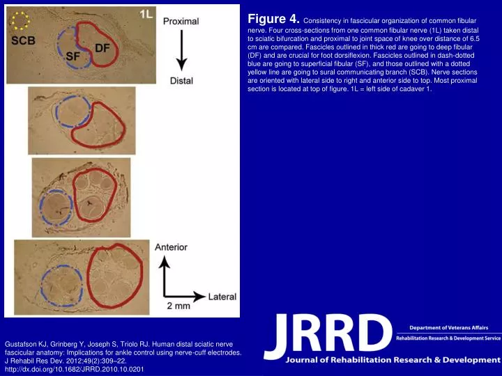
Figure 4. Consistency in fascicular organization of common fibular nerve. Four cross-sections from one common fibular nerve (1L) taken distal to sciatic bifurcation and proximal to joint space of knee over distance of 6.5 cm are compared. Fascicles outlined in thick red are going to deep fibular (DF) and are crucial for foot dorsiflexion. Fascicles outlined in dash-dotted blue are going to superficial fibular (SF), and those outlined with a dotted yellow line are going to sural communicating branch (SCB). Nerve sections are oriented with lateral side to right and anterior side to top. Most proximal section is located at top of figure. 1L = left side of cadaver 1. Gustafson KJ, Grinberg Y, Joseph S, Triolo RJ. Human distal sciatic nerve fascicular anatomy: Implications for ankle control using nerve-cuff electrodes. J Rehabil Res Dev. 2012;49(2):309–22.http://dx.doi.org/10.1682/JRRD.2010.10.0201