Download

1 / 24
240 likes | 1.14k Vues
Survival guide. Peds Soft Tissue Neck Xrays. Approach alignment bones -- vertebral bodies cartilage -- disc spaces C1 and C2 positioning of the neck pre-vertebral space epiglottis subglottic space Needs to be in extension preferably at end-inspiration.

E N D
Survival guide Peds Soft Tissue Neck Xrays
Approach alignment bones -- vertebral bodies cartilage -- disc spaces C1 and C2 positioning of the neck pre-vertebral space epiglottis subglottic space Needs to be in extension preferably at end-inspiration The Soft-Tissue Lateral neck Film
Micro GAS, staph aureus, anaerobes Complications UA obstruction pus or secretion aspiration mediastinitis sepsis dehydration Retropharyngeal abcess
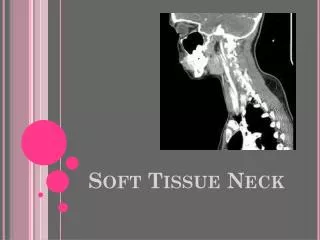
Epiglottitis:Xray appearance • ‘thumb-like’ appearance of epiglottis • thickened aryepiglottis folds • loss of normal pre-epiglottic (vallecular) space
Management minimal agitation airway maintenance IV antibiotics IV hydration analgesia blood and epiglottic cultures Micro staph. Aureus and GAS most common also strep. pnemoniae Hib prior to vaccination Epiglottits
Most common upper airway obstruction in children, peak at 2 yrs Parainfluenza types 1 and 2, influenza A and B, rhinovirus edema of subglottic space worse during late night and early morning Croup (laryngotracheobronchitis)
Bacterial Tracheitis • Rare complication of viral croup • 6mo – 8yrs, mean age 5 yrs • S. aureus, S. pneumo, Group A strep, H. flu, M. catarrhalis • Best diagnosed by bronchoscopy – thick inflammatory exudate with sloughed mucosa in lumen • Lateral neck x ray: hazy tracheal air column with luminal soft tissue irregularities • 55 - 80% patients require intubation +/or tracheostomy • Cefuroxime 50mg/kg IV Q8H +/- endotracheal suctioning prn