Download
1 / 21
270 likes | 977 Vues
Detection of Prion Diseases. Amir Imani, Danish Tanwar, Rana Khafagy, Marwa Saad . PHM142 Fall 2012 Coordinator: Dr. Jeffrey Henderson Instructor: Dr. David Hampson. What We ’ ll be Discussing. Prion Diseases: Background Guaranteed Method of Detection Genetic Screening

E N D
Detection of Prion Diseases Amir Imani, Danish Tanwar, Rana Khafagy, Marwa Saad PHM142 Fall 2012 Coordinator: Dr. Jeffrey Henderson Instructor: Dr. David Hampson
What We’ll be Discussing • Prion Diseases: Background • Guaranteed Method of Detection • Genetic Screening • Neuroimaging Techniques • Blood Test • EEG • Lumbar Puncture • Surround Optical Fiber Immunoassay (SOFIA) • RT-QuIC • Future Developments
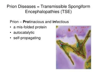
Prion Diseases: Background • Prion diseases are known as Transmissible Spongiform Encephalopathies (TSEs) • These conditions are progressive, degenerative diseases that affect the brain and central nervous systems • Prion diseases result in four physiological effects: 1. Spongiform change (holes in the brain matter) 2. Neural loss 3. Astrocytosis (increase in astrocytes because of the loss of neurons) 4. Amyloid plaques of prions
Prion Diseases: Background • Prion diseases are difficult to identify • They are often mistaken for other neurodegenerative conditions such as Alzheimer’s • There are 4 main classes of TSEs in humans (though there are variations within them) • Detection methods can vary based on the condition and TSEs can either be genetic or contracted (typically through contaminated brain matter)
Guaranteed Method of Detection • Analyzing brain samples is the only way to confirm the presence of a prion disease (though a proper diagnosis can be done with other means) • This is done through either an autopsy or a brain biopsy • The samples are studied to see if they exhibit the typical damage and have amyloid plaques of prions caused by TSEs. Brain Atrophy caused by Creutzfeldt–Jakob disease The “Spongiform” effect of prion diseases
Genetic Screening • Not all prion diseases have a genetic cause; however, mutations in the PRNP gene can result in prion disease • PRNP (PRioNProtein) gene is responsible for the creation of normal Prion Proteins • Every TSE that has a genetic link has been shown to involve a mutation in the PRNP gene • It is inherited in an autosomal dominant manner (located on chromosome 20) • Healthy prion proteins are not problematic and are predominantly expressed in the brain and CNS • There is no definitive evidence of their role, but they are thought to be involved in circadian rhythm, among a host of other uses
Genetic Screening • Some of the PRNP mutations that lead to TSEs include: • Genetic screening involves a basic sequence analysis of the coding and flanking sections of the PRNP gene. • Note: Though genetic mutations can lead to these conditions, it does not mean they are the main cause (For example: Only 5-10% of CJD cases are caused by a genetic mutation).
Neuroimaging Techniques • Spongiform change and plaque deposits can be detected early through a variety of imaging studies: • Computerized Tomography (CT) • Magnetic Resonance Imaging (MRI) • Electroencephalogram (EEG) • CT Scan has gradually been replaced by MRI • Atrophy of brain tissues is often seen • High intensity signals: a sign of cognitive defects • Drawback: False Positive: The brain biopsy may indicate the presence of other diseases instead, such as Alzheimer’s MRI Scan of a patient diagnosed with Creutzfeldt-Jakob Disease (CJD)
Blood Test • Used to distinguish TSEs from other diseases • But it is NOT applicable to all TSEs • Much more sensitive than previous methods
Breakthrough in Detecting Prion Infections • A solid matrix developed to capture disease-associated prion proteins • Immunodetection carried out Results: • 15 out of 21 samples were correctly identified up by the assay Future Research: • Research required to disprove the possibility of false positive results • Further studies needed to assess the long-term effects
Electroencephalogram (EEG) • Measure electrical activity of the brain • Electrodes placed all over scalp to record impulses of neurons • Used to diagnose CJD 8 to 12 weeks after onset of symptoms • Periodic sharp wave complexes (PSWCs) indicate progressive state of CJD Rules out Alzheimer’s • Shows synchronized, sharp waves that are very specific to common CJD • Disadvantages: Kuru, FFI, and other forms of CJD do not have predictable EEG patterns
Lumbar Puncture • Collect CSF for analysis • Patient placed in fetal position anesthetic given spinal needle pushed into lower back 2 pushes Remove needle while applying pressure on puncture site • Sample analyzed for 14-3-3 protein (released from damaged or dying nerve cells) and tau protein • 14-3-3 detectable in 90% of all CJD • Excludes brain infections, viral encephalitis, and Alzheimer’s • Disadvantage: Cannot detect PrP-Sc disease + Nonspecific
Surround Optical Fiber Immunoassay (SOFIA) • Captures fluorescent emissions from blood or urine • Detects even the smallest amount of PrPSC • Very sensitive and dynamic • High levels of detection • Disadvantage: • Time consuming and expensive • Need experts for interpretation
Surround Optical Fiber Immunoassay (SOFIA) Figure: Intensity peaks using Protein Misfolding Cyclic Amplification (PMCA) technology to distinguish PrPC from PrPSC
RT-QuIC • in vitro shaking helps to accelerate the reaction, which enables the assay to produce results more quickly
RT-QuIC • Inverse relationship between protein concentration and fibril formation • Protein is induced in the presence of denaturant or low pH leading to increased oligomer formation
RT-QuIC • Tested 18 people with CJD and 35 people with other neurodegenerative diseases • CJD was correctly diagnosed more than 83% of the time and no false positives were produced • This technique allows for ante-mortem confirmation of Prion diseases • Sensitive: Can detect sub-femtogram amount of PrPSc in hamsters within a single day • Specific: Specific for prion disease; does not give false positives Alzheimer’s disease • Speedy: Yields results within 48 hours
Prion Disease: The Future? • Clinical implications: Can RT-QuIC methods be replicated in a larger sample? • Can this method be used to detect other prion diseases? • Can RT-QuIC methods be used to screen donated blood?
Summary • Analyzing brain samples is the only way to confirm the presence of a prion disease. This is done through either an autopsy or a brain biopsy. • Neuroimaging techniques can lead to false positives. • EEG only accurate at detecting common CJD by looking at distinct sharp peaks using PSWCs • Lumbar puncture can detect many variations of CJD accurately by analyzing 14-3-3 and tau proteins in cerebrospinal fluid • SOFIA detects low concentrations of PrPSC, leading to accurate diagnosis • RT-QuIC is able to detect prion disease ante-mortem.
References • Atarashi, R., Sano, K., Satoh, K., Nishida, N. (2011). Real-time quaking-induced conversion: a highly sensitive assay for prion detection. Prion, 5(3), 150-153. • Chang, B. G., Gray, P., Piltch, M., Bulgin, M. S., Sorensen-Melson, S., Miller, M. W., Davies, P., Brown, D. R., Coughlin, • D. R., & Rubenstein, R. (2009). Surround optical fiber immunoassay (SOFIA): An ultra-sensitive assay for prion protein detection. Journal of Virological Methods, 159 (1), 15-22. • Collins, S., McLean, C.A., Masters ,C.L. (2001). Gerstmann-Straussler-Scheinker syndrome,fatal familial insomnia, and kuru: a review of these less common human transmissible spongiform encephalopathies. J Clin Neurosci, 8 (5), 387–97. • Creutzfeldt-Jakob Disease Foundation. (2007). Creutzfeldt-Jakob Disease and other prion diseases, 1-36. • Edgeworth, J.A., Farmer, M., Sicilia, A., Tavares, P., Beck, J., Campbell, T., Lowe, J., Mead, S., Rudge, P., Collinge, J., Jackson, G.S. (2011). Detection of prion infection in variant Creutzfeldt-Jakob disease: a blood-based assay. The Lancet. 377, 487-493. • Kallenber, K., Schulz-Schaeffer, W.J., Jastrow, U., Poser, S., Metssner, B., Tschampa, H.J., Zerr, I., Knauth, M. (2006). Creutzfeldt-Jakob Disease: Comparative Analysis of MR Imaging Sequences. AJNR Am J Neuroradiol. 27, 1459-1462. • Macfarlene, R.G., Wroe, S.J., Collinge, J., Yousry, T.A., Jager, H.R. (2007). Neuroimaging findings in human prion disease. J Neurol Neurosurg Psychiatry. (78), 664-670. • Mastrianni, J. A. (2003). Genetic Prion Diseases in Gene Reviews. Seattle, Washington: GeneReviews. • Montagna, P., Gambetti, P., Cortelli, P., Lugaresi, E. (2003). Familial and sporadic fatal insomnia. Lancet Neurol, 2 (3), 167–76. • University of California San Francisco. Primer of Human Prion Disease. Department of Neurology. Retrieved on November 6, 2012 http://memory.ucsf.edu/sites/all/files/download/MAC_RPD_Primer.pdf. • Wang, P.S., Wu, Y.T., Hung, C.I., Kwan, S.Y., Teng, S., & Soong, B.W. (2008). Early detection of periodic sharp wave complexes on EEG by independent component analysis in patients with Creutzfeldt-Jakob disease. Journal of Clinical Neurophysiology, 25 (1), 25-31. • Woo, L. J., & Khoshbin, S. (2008). Clinical neurophysiology and electroencephalography. Massachusetts General Hospital Comprehensive Clinical Psychiatry, 12 (8), 75-87. • Zomosa-Signoret, V., Arnaud J.D., Fontes, P., Alvarez-Martinez, M.T., Liautard, J.P. (2008). Physiological role of the cellular prion protein. Vet. Res. 39 (4), 9.