Download
1 / 33
340 likes | 542 Vues
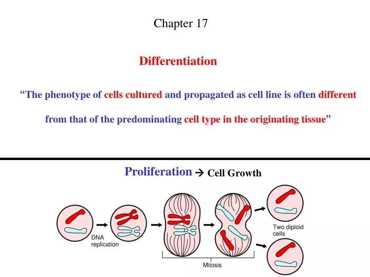
Chapter 17. Differentiation. “ The phenotype of cells cultured and propagated as cell line is often different from that of the predominating cell type in the originating tissue ”. Proliferation. Cell Growth. Proliferation and Differentiation.

E N D
Chapter 17 Differentiation “The phenotype of cells cultured and propagated as cell line is often differentfrom that of the predominating cell type in the originating tissue” Proliferation Cell Growth
Proliferation andDifferentiation “Cell proliferation is incompatible with the expression ofdifferentiated properties” Malignant tumor cells: sometimes break the restriction Melanin continues to be synthesized while the cells areproliferating
Differentiation (1) “The process leading to the expression of phenotypic propertiescharacteristics pf the functionally mature cell in vivo” (2) “The combination of Constitutive and Adaptive propertiesfound in the mature cell” Constitutive: stably expressed w/o inductionAdaptive: with positive or negative regulation of expression
produce extracellular matrix • express the hematopoietic cell surface markers (CD34+ & CD45+)and fibroblast marker (collagen) • migrate to wound sites (wound healing) Terminal Differentiation “A cell has progressed down a particular lineage to a point, thatthe cell cannot progress anymore” Examples: Neurons, Skeletal Muscle Cells, Keratinocyte Exclusion: Fibrocytes
Two main pathways to Differentiation 1. In constantly renewing tissues:--e.g., epidermis, intestinal mucosa, blood--Pluripotent undifferentiated stem cells Unipotent Progenitor cells Terminal differentiated cells--this process gives rise to mature, differentiated cells that normal will not divide 2. In tissues that do not turn over rapidly:--e.g., fibrocytes, blood vessel endothelial cells, glial cells, hepatocytes, satellite cells-- Cells lose their differentiated properties and reenter the cell cycle-- When the tissue has regained the appropriate cell density by division,cell proliferation stops and differentiation is reinduced.--This type of renewal is rapid
Whether the cells that reenter the cell cycle to regenerate the tissue are phenotypically identical to the bulk of thedifferentiated cell population ??
Dedifferentiation Cells in cell culture can lose properties they originally had, such as protein expression, or change shape When Dedifferentiation occurs: 1. Adaptive Process: regaining the differentiated phenotype under the right inducers 2. Selective Process: because of cell’s greater proliferative potential
Reversible Cell Properties • Irreversible characteristic: e.g., mature keratinocyte-- cessation of DNA synthesis • Reversible characteristic: e.g., hepatocyte-- re-induction of albumin synthesis Stem cells Progenitor cells Differentiated cells irreversible irreversible Differentiation Process
Commitment & Cell Lineage • A hematopoietic stem cell, after commitment to lymphocytic differentiation,would not change lineage at a later stage • Stem cell Progenitor cell Differentiated cell • Someprogenitor cells can revert to stem cells with multilineage potential such irreversible commitment must occur much later than “the point” • Most cultures are derived from normal tissues (stem cells, late progenitor cells,differentiated cells) do not alter to a different lineage • Tumor culture: changes its commitment K562 cell line: isolated from a myeloid leukemia & be capable of erythroid differentiation C6 glioma of rat: expresses both astrocytic & oilgodendrocytic features • Cell lines are regarded as a mixed population of stem cells, progenitor cells, anddifferentiated cells
Re-Differentiation (I) • The majority of cell lines do not express fully differentiated properties • Developing Organ Culture System:Advantages:(1) retain three-dimensional, high-cell-density tissue architecture(2) prevent dissociation(3) prevent selective overgrowth of undifferentiated cellsDisadvantages:(1) inability to propagate large numbers of cells(2) heterogeneity of the tissue sample • In pure populations of cells:--to reinduce the differentiated phenotype by recreating the correct environment--to define individual influences exerted on the induction and maintenance of differentiation
Glial cells: oligodendrocyte & astrocyte CNS Schwann cells ?? Schwann cells:1. using cholera toxin as a mitogen 2. optimal condition: glial growth factor + forskolin + 10% serum
Re-Differentiation (II) Environment (I) • Brain (O2A common progenitor cell of oligodendrocyte and progenitor cell oftype 2 astrocyte)-- in the mixture of PDGF and bFGF: remains a proliferating progenitor cell-- in the absence of growth factor or serum: differentiate into an oilgodendrocyte--in the fetal bovine serum or a combination of ciliary neurotropic factor (CNTF) & bFGF differentiate into a type 2 astrocyte • Cardiac muscle cell:-- in serum and bFGF: remains undifferentiated and proliferative properties-- in the absence of serum: differentiate
Re-Differentiation (II) Environment (II) 3. MDCK cell (cell line from dog kidney) / salivary gland epithelium / mammary epithelium: --in the mixture of epimorphin and HGF (hepatocyte growth factor) inducing tubule formation (differentiation) 4. Prostatic epithelium / epidermal epithelium: -- in the presence of KGF (keratinocyte growth factor): differentiation 5. Primitive embryonal stem (ES) cell: --differentiate Spontaneously --in bFGF & SCF (stem cell factor) & LIF (lymphocyte inhibitory factor) kept in the undifferentiated proliferative phase
Stem Cell Plasticity • Unipotent stem cell: e.g., stem cell in the basal layer of dermis-- will give rise to only one lineage-- differentiate into a keratinocyte • Bipotent stem cell: e.g., a lymphoid stem cell-- will give rise to two lineages-- differentiate into T- or B-lymphocyte • Multipotent stem cell: e.g., stem cell in the bone marrow-- will give rise to more than two lineages-- differentiate into granulocytes, monocytes, megakaryocytes, mast cells, erythrocytes • Totipotent stem cell: e.g., embryonal stem cell (ES cell)-- will give rise to every cell lineages
Stem Cell • Non-regenerative tissues do have stem cells-- Brain (Neurons) • Tissue localization does not necessarily mean lineage commitmentand reduced potency-- Liver stem cells can generate neurons-- Bone marrow stem cells can generate cardiac muscle cell, hepatocytes, neurons-- Muscle stem cells can generate hematopoietic cells-- Neural stem cells can generate endothelial cells • Third source:Umbilical cord:--hematopoietic, multipotency stem cells--fewer ethical limitations than human ES cells--greater longevity in the cell line generated
Markers of Differentiation 1. Markers of the mature phenotype representing terminal differentiation 2. Examples: -- Epithelium: cytokeratins -- Astrocytes: glial fibrillary acidic protein -- Erythrocyte: hemoglobin -- Hepatocyte:albumin -- Keratinocytes: transglutaminase or involucrin -- Oligodendrocyte: glycerol phosphate dehydrogenase 3. Looking for the expression of differentiation marker proteins: -- RT-PCR (?): will not necessarily confirm synthesis of proteins[to distinguish between low levels and high levels of gene expression] -- cDNA Microarray (?) -- Western blot -- Immunofluorescent staining (Flowcytometry or Fluorescent microscopy) -- 2D gel (?)
Regulation of Differentiation (4) (1) (3) (5) (2)
Induction of Differentiation Cell Interaction • Heterotypic Cell Interaction:--is responsible for initiating and promoting differentiation--mutual interaction between cells originating different germ layers (endoderm, mesoderm, ectoderm) promote differentiation Keratinocyte Fibroblasts
Reciprocal Paracrine Interaction: for epidermal maturation Autocrine factor Homotypic paracrine factor kerotinocyte Heterotypic paracrine factor fibroblasts
Induction of Differentiation Cell Interaction 2. Homotypic Cell Interaction:-- Gap junction communication: e.g., cAMP, Ca2+, diacylglycerol-- Electrical charge -- harmonizes / balances the expression of differentiation, rather than initiating its expression
Induction of Differentiation Systemic factors / Physiological inducersorExogenous factors / Nonphysiological inducers Physiological inducers • Hormones: hydrocortisone / glucagon / thyroxin • Vitamins: retinoids (endothelium) / vitamin D3 • Inorganic ions: calcium (keratinocyte)
Induction of Differentiation Systemic factors / Physiological inducersorExogenous factors / Nonphysiological inducers Nonphysiological inducers • Planar-polar compounds: DMSO / HMBA • Cytotoxic drugs: genistein / mitomycin C • Signal transduciton modifiers: PMA
Dimethyl sulfoxide (DMSO) • mouse erythroleukemia / neuroblastoma / myeloma / mammary carcinoma response to DMSO by differentiating • The action of DMSO is unclear !!! • Possible mechanisms:-- by changes in membrane fluidity-- by influence on protein kinase C or phospholipase D-- by alternations in DNA methylation or histone acetylation • The induction of differentiation by DMSO may be phenotypically normal
Induction of Differentiation Cell-Matrix Interaction Matrix-induced differentiation • liver-derived matrix material: induced expression of the albumin gene in hepatocyte • Collagen: essential for the functional expression of epithelial cells essential for endothelial cells to mature into capillaries • Synthetic matrix (poly-D-lysine): promote neurite extension in neuronal culture • Matrigel (laminin+collagen+proteoglycans):endothelial cells & epithelial cells differentiate effectively • Modulation of growth factor activity
Neurite extension in neuronal culture www.actaps.com.cn/.../htmlwenzhang/2006-6-06.htm
Heparan Sulfate: cell surface proteoglycan http://imbs.massey.ac.nz/Staff/norris.html 1. Transmembrane heparan sulfate proteo-glycans (HSPGs) may acts as low-affinitygrowth factor receptor 2. HSPGs may transport these growth factorsto high-affinity receptors http://www.med.unibs.it/~airc/hspgs.html
Induction of Differentiation Cell shape (Polarity) • Hepatocytes:-- flattened shape-- Cuboidal or columnar shape • Thyroid epithelium: in a filter well assembly--lower (basal) surface: released triiodothyronine--upper (apical) surface: released thyroglobulin
Induction of Differentiation Oxygen Tension http://www.mattek.com/pages/in_vitro_basics Air/Liquid Interface (ALI)
Induction of Differentiation Oxygen Tension • Positioning of epidermal cells at the ALI:-- fully keratinized squamous differentiation • Location of alveolar type II cells at the ALI:-- for optimal differentiation • Location of tracheal epithelium at the ALI:-- become mucus secreting • Positioning of tracheal epithelium at the bottom of the dish:-- become squamous • ALI: -- enhances gas exchange-- w/o risking free radical toxicity
Differentiation & Malignancy • Cancer: a failure of cells to differentiate normally • With increasing progressing of cancer:-- histology of a cancer indicates poorer differentiation • Patients with poorly differentiated tumors:-- have a lower survival rate than patients with differentiated tumors • Many tumors grown in tissue culture can be induced to differentiate