Download
1 / 45
530 likes | 1.37k Vues
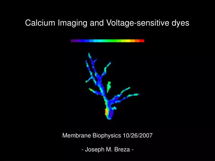
Calcium Imaging and Voltage-sensitive dyes. Membrane Biophysics 10/26/2007. - Joseph M. Breza -. Outline. 1) Backgroud Calcium-sensitive dyes, Calcium imaging.

E N D
Calcium Imaging and Voltage-sensitive dyes Membrane Biophysics 10/26/2007 - Joseph M. Breza -
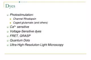
Outline 1) Backgroud Calcium-sensitive dyes, Calcium imaging 2) Riera CE, Vogel H, Simon SA, le Coutre J. Artificial sweeteners and salts producing a metallic taste sensation activate TRPV1 receptors, Am J Physiol Regulatory Integrative Comp Physiol 293:626-634, 2007. 3) Background Voltage-sensitive dyes, imaging
Calcium The ion of the nervous system Neurotransmitter release into synapse via vesicle release proteins Signaling cascades Cell division Blood clotting Fertilization Amount of Calcium needed for effect is relatively small
Fluorescence: Is the phenomenon in which the molecular absorption of a photon triggers the emission of another photon with a longer wavelength. (named after calcium fluoride “Fluorite”) * Stimulate in the ultraviolet range, and the emitted light is in the visible range.*
Agar Plate of Fluorescent Bacteria Colonies QUIN 2 was a member of the first generation of Ca2+ chelators introduced by Tsien in 1980
Calcium Imaging Fluorescence detection system 1) Fluorophore- fluorescent molecule (photo bleaching) 2) Wavelength filters 3) Excitation source (light source UV) 4) Detector Fluorescence instruments 1) Spectrofluorometer – analyzes fluorescence (before – after) 2) Flow cytometer- analyzes, sorts, counts microscopic particles in stream of fluid-doesn’t produce and image of the cell 3) Fluorescence microscope - filters weak fluorescent light from other lights
Calcium sensitive dyes are Chelators Many different types of calcium fluorescent dyes Type used depends on the cell type - experience Research goal (salt/lipophilic forms, low/intermediate/high affinity, fast calcium spikes, fluorescence, method of delivery, etc) Low-affinity calcium indicators Fura-FF BTC Fura-2 Fura-5, Indo-1) Excited by UV light Intermediate-affinity calcium Fura-4F Fura-5F Fura-6F Excited by UV light Excited by visible light under scanning laser confocal microscopy High-affinity and selectivity (BAPTA) Calcium Green, Calcium Orange – Tomchik et al 2007
Fluorescent microscope Protects eyes from UV light
Figure 2 PLCβ2 negative G-proteins CaGD- Calcium Fluorescence
Figure 4 PLCβ2-GFP Mouse (Receptor cell) GAD-GFP Mouse (Presynaptic cell)
Artificial sweeteners and salts producing a metallic taste sensation activate TRPV1 receptors Am J Physiol Regulatory Integrative Comp Physiol 293:626-634, 2007. Authors: Riera CE, Vogel H, Simon SA, le Coutre J.
Voltage-sensitive dyes A plethora of them to choose from…depends on tissue (experience) Vary considerably in active duration, loading, etc. “Sticks” to the plasma membrane Changes in membrane potential are recorded with a photo diode (464 pixels 1010 photons per frame) ~1nA photocurrent and dye sensitivity of ~1%/100mV Can calculate membrane potential within 3 mV Change in Fluorescence can be between ~1-21% / 100mV change (RH421).
Measure Active Conductances – Voltage gated Na, K, Ca2+ Channels What’s the advantage?
Optical recording of synaptic release Digital Camera Pseudocolor output of camera Transgenic olfactory bulb
The pH-sensitive protein: synaptopHluorin Mutagenized Green Fluorescence Protein Neuron. 2005 Dec 22;48(6):1039-53. can accept a proton to change fluorescence fused to VAMP2 expressed in mice targets to synaptic vessicles in vivo When synaptic vessicle fuses with membrane pH increase almost instantly fluorescence increases almost instantly
pH of: vesicle extracellular Gero Miesenböck, Dino A. De Angelis and James E. Rothman Nature 394, 192-195(9 July 1998)
Odorant Representations Are Modulated by Intra- but Not Interglomerular Presynaptic Inhibition of Olfactory Sensory Neurons McGann JP, Pírez N, Gainey MA, Muratore C, Elias AS, Wachowiak M. Question: How does presynaptic inhibition really work in the olfactory bulb glomerulus?
Single glomerulus: 2 bundles of OSNs Single bundle are cumulative Paired-pulse results in depression Depression alleviated by: GLU blockers GABA blockers
Optical recording of voltage changes in vivo photodiode array Pseudocolor output of the array turtle http://www.redshirtimaging.com
Voltage-sensitive dyes A plethora of them to choose from…depends on tissue (experience) Vary considerably in active duration, loading, etc. “Sticks” to the plasma membrane Changes in membrane potential are recorded with a photo diode (464 pixels 1010 photons per frame) ~1nA photocurrent and dye sensitivity of ~1%/100mV can calculate membrane Potential within 3 mV Change in Fluorescence can be between ~1-21% / 100mV change (RH421).
Binaral interaction and centrifugal input enhances spatial contrast in olfactory bulb activation. Singer, Benjamin H., Kim, Soyoun & Zochowski, Michal European Journal of Neuroscience 25 (2), 576-586. Question: What role do centrifugal inputs play in OB activation?
Schematic of olfactory bulb neurotransmitters and cellular connections Note: All of these different neurotransmitters are coming from BOTH sides of brain Doty RL (1995), Handbook of Olfaction and Gustation, Marcel Dekker.
Experimental design and controls Replicates with one odorant
Paired-pulse with different odorants results in increased inhibition This would have been difficult to see with standard electrophysiology or behavior
Flash-activation of compounds won the 1967 prize in Noble Prize in Chemistry Light-induced isomerization acetylcholine agonists by Wasserman and Erlanger 1971 only trans isomer biologically active (Bis-Q) Many kinds today used on: Ca++ cNMP glutamate GABA peptides
Photolysis of Caged Glutamate UV photon Caging compound Glutamate Requirements of caging compounds/ systems: Masks the biomolecule completely Releases the biomolecule completely No deleterious by-products No biomolecule breakdown http://flavor.monell.org/%7Eloweg/Techniques.htm
Photolysis and Recording Set-up Beam can be moved in X-Y plane and in Z-plane for discrete anatomical placement Or discrete channels can be placed with the compound http://flavor.monell.org/%7Eloweg/Techniques.htm
Discrete Release of Caged Compound Lowe, G. J Neurophysiol 88: 64-85 2002
Flash photolysis reveals a diversity of ionotropic glutamate receptors on the mitral cell somatodendritic membrane. Lowe G. J Neurophysiol. 2003 90(3):1737-46. Question: Are glutamate rececptors located on the mitral cell soma/ proximal dendrites?
Glutamate elicts fast and slow currents Photolysis at arrow on soma/ proximal dendrite NBQX= AMPA antagonist APV= NMDA antagonist (comp) dCK= NMDA glycine site antagonist (comp) Mg++ -free bath Lowe, G. J Neurophysiol 90: 1737-1746 2003; doi:10.1152/jn.00180.2003
Competitive inhibition of NMDA current Lowe, G. J Neurophysiol 90: 1737-1746 2003; doi:10.1152/jn.00180.2003
Fast Glutamate Current = AMPA? All slow glutamate currents blocked Fast current remains CTZ= AMPA postitve allosteric modulator Lowe, G. J Neurophysiol 90: 1737-1746 2003; doi:10.1152/jn.00180.2003
Fast Glutamate Current = a little Kainate? All slow glutamate currents blocked Fast current remains SYM2206= Kainate blocker