Download
1 / 29
330 likes | 567 Vues
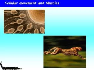
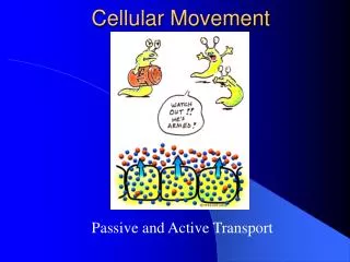
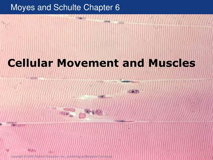
Cellular Movement and Muscles. Cellular movement. Movement is a property of all cells. Some cells (such as this amoeba) can move through their environment. All cells can move components through the cytoplasm (such as the vesicles in this amoeba). Cytoskeleton and Motor Proteins.

E N D
Cellular movement Movement is a property of all cells Some cells (such as this amoeba) can move through their environment All cells can move components through the cytoplasm (such as the vesicles in this amoeba)
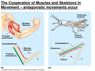
Cytoskeleton and Motor Proteins • All physiological processes depend on movement • Intracellular transport, changes in cell shape, cell motility, and animal locomotion • All movement is due to the same machinery • Cytoskeleton – protein-based intracellular network • Motor proteins – enzymes that use energy from ATP
Cytokeleton Composed of actin and microtubules Fluorescently labeled cell Actin – red Microtubules – green Nuclei - blue
The Cytoskeleton and Movement • Three ways to use the cytoskeleton for movement • Cytoskeleton roadway and motor protein carriers • Reorganization of the cytoskeletal network • Motor proteins pull on the cytoskeletal rope Figure 6.1
Microtubules • Tube-like polymers of tubulin • Organized into many arrangements • Anchored near the nucleus and the plasma membrane • Microtubule-organization center (MTOC) (-) • Integral proteins (+) Figure 6.2
Microtubule Structure • Polymers composed of the protein tubulin • Dimer of a–tubulin and b-tubulin Figure 6.4
Microtubules Composition and Formation • Microtubules have a plus and minus end Figure 6.5
Microtubules • Minus end of the microtubule is anchored at the Microtubule-organization center (MTOC) • Plus end of the microtubules anchored by Integral membrane proteins at the plasma membrane Figure 6.2
Microtubules can grow and shrink A microtubule can grow or shrink from either end “Dynamic Instability” Fig 6.6a
Factors affecting Dynamic Instability • Local concentration of tubulin affects microtubule dynamics Figure 6.6
Microtubule dynamics regulated by MAPs MAPs: Microtubule associate proteins Bind to microtubulues and stabilize or destabilize structure Figure 6.7
Motor proteins Motor proteins can move along microtubules • alpha-Tubulin: pale blue • beta-Tubulin is pale green • Kinesin walks towards the plus-end of microtubules (right side of picture) Hoenger, A., Thormählen, M., Diaz-Avalos, R., Doerhoefer, M., Goldie, K.N., Müller, J. and Mandelkow, E. (2000) A new look at the microtubule binding patterns of dimeric kinesins. J Mol Biol, 297, 1087-103.
Movement Along Microtubules • Direction is determined by polarity and the type of motor protein • Kinesin move in + direction • Dynein moves in – direction • Fueled by ATP • Rate of movement is determined by the ATPase domain of the protein and regulatory proteins • Dynein is larger than kinesin and moves 5-times faster
Microtubule Functions • Move subcellular components • e.g., Rapid change in skin color Figure 6.3
Vesicle Traffic in a Neuron Figure 6.8
Microtubule function - Cilia and Flagella • Cilia – numerous, wavelike motion • Flagella – single or in pairs, whiplike movement • Composed of microtubules • Arranged into axoneme • Movement results from asymmetric activation of dynein Figure 6.9
Microtubules and Physiology Table 6.1
Microfilaments • Other type of cytoskeletal fiber • Polymers composed of the protein actin • Often use the motor protein myosin • Found in all eukaryotic cells • Movement arises from • Actin polymerization • Sliding filament model using myosin (more common)
Actin filament Structure and Growth • Polymers of G-actin called F-actin • Spontaneous growth (6-10X faster at + end) • Treadmilling when length is constant • Capping proteins increase length by stabilizing minus end Figure 6.10
Microfilament Arrangement Figure 6.11
Actin Polymerization • Amoeboid movement • Two types • Filapodia are rodlike extensions • Neural connections • Microvilli of digestive epithelia • Lamellapodia resemble pseudopodia • Leukocytes • Macrophages Figure 6.12
Myosin – a motor protein • Motor protein that works with actin filaments • Most common type of movement • Myosin is an ATPase • Converts energy from ATP to mechanical energy • 17 classes of myosin with • multiple isoforms • Similar structure • Head, tail, and neck Figure 6.14
Myosin as a motor protein Myosin moves along actin • Analogous to pulling yourself along a rope • Actin: the rope • Myosin: your arm
Sliding Filament model Figure 6.15
Sliding Filament Model. • Two processes • Chemical • Myosin binds to actin (Cross-bridge) • Structural • Myosin bends (Power stroke) • Cross-bridge cycle • Formation of cross-bridge, power stroke, and release • Need ATP to attach and release Figure 6.15
Variation in myosin function • Two factors • Unitary displacement • Distance myosin steps during each cross-bridge cycle • Depends on • Myosin neck length • Myosin placement (helical structure of actin) • Duty cycle • Cross-bridge time/cross-bridge cycle time • Typically 0.5 • Use multiple myosin dimers to maintain contact Figure 6.16
Sliding filament assay Figure 18-22, Lodish 4th edition. The sliding-filament assay
Actin and Myosin Function Muscle contraction Table 6.2