Download
1 / 46
460 likes | 643 Vues
2 ND ANNUAL RAPID REVIEW 12.O3.12 CARE OF PATIENT WITH CHEST INJURIES. Sujitha .E, Lecturer, Faculty of Nursing, Sri Ramachandra University, Porur. Chest cavity. Soft tissues Lungs Heart Great vessels diaphragm oesophagus. Bony areas. Ribs Sternum Clavicle Tracheo broncheal

E N D
2ND ANNUAL RAPID REVIEW 12.O3.12CARE OF PATIENT WITH CHEST INJURIES Sujitha .E, Lecturer, Faculty of Nursing, Sri Ramachandra University, Porur
Chest cavity Soft tissues • Lungs • Heart • Great vessels • diaphragm • oesophagus
Bony areas • Ribs • Sternum • Clavicle • Tracheo broncheal tree
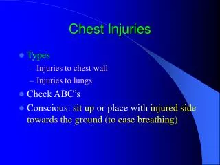
Classification Blunt injuries Penetrating injuries
Etiology • Motor vehicle accidents • Fall from height • Violence • Iatrogenic
Mechanisms involved • Acceleration force • Deceleration force • Transmission of blunt internal force to force to structures • Direct trauma • Compression
Chest trauma • Chest wall injuries • Sternal fractures • Flail chest • Pulmonary and pleural injuries • Traumatic asphyxia • Tracheo bronchial injuries • Pneumothorax • Hemothorax • Mediastinal injuries • cardiac injuries • Great vessel injuries • Diaphragmatic injuries • Oesophageal injuries
Pulmonary injuries Pneumothorax • Collection of air in the space between the parietal and visceral pleura
Tension pneumothorax An expanding collection of intra pleural air without communication with external environment • Clinical manifestations • Distended neck veins • Hypotension/hypoperfusion • Absent breath sounds on affected side • Tracheal deviation to contra lateral side
Management • Immediate needle aspiration • 14 gauge IV needle of length more than 4.5 cm and catheter into pleural space through chest wall in MCL at second intercostal space(temporary measure) • Large bore chest tube thoracostomy
Open pneumothorax (sucking chest wound) A communication between the pleural space and surrounding atmospheric pressure Respiration is the function of negative pressure inside the thoracic cavity , positive atmospheric pressure and elastic recoil of lungs
Pneumothorax • Clinical manifestations • Air entry and breath sounds diminished in the affected side • Impaired chest wall motion
Pathophysiology Negative intrapleural pressure during inspiration Air leak into the pleural cavity Increased intra thoracic pressure Reduced vital capacity and venous return
Pneumothorax • Diagnosis • Chest radiography(double pleural markings) • Ultrasound • Management • Cover the wound with a three sided dressing • Air can escape during expiration but do not enter during inspiration(one way valve) • Chest tube insertion
Open pneumothorax 3-side dressing Asherman chest seal
Massive hemothorax Accumulation of at least 1500 ml or two thirds of the available hemithorax in an adult
Hemothorax • Life threatening by three mechanisms • Acute hypovolemia causing decreased preload • Collapsed lung promoting hypoxia • Hemothorax compressing venacava • impairing preload
Hemothorax • Clinical manifestations Abnormal vital signs Dullness to percussion Diminished breath sounds • Diagnosis Plain chest radiography completely opacifiedhemithorax Ultrasonography-fluid between chest wall and lung
Management • Chest tube insertion Care of chest tube • Position-last hole 2.5-5 cm inside chest wall • Suction chamber with 20-30 cm of water • Never clamp the tubes • Bottle at 1-2 ft lower than patient’s chest • Left in place for 24 hrs after leak has stopped
Flail chest • Free floating lung segment that is no longer connected to the rest of the thorax • Cause • Segmental rib fractures in two or more locations of the same rib of three or more adjacent ribs
Flail chest • Clinical manifestations • Paradoxical inward movement of the involved portion of the chest wall during inspiration and outward movement during expiration
Pathophysiology-flail chest Decreased ventilatory efficiency Increased work of breathing Hypoxemia Sudden respiratory arrest
Management-Flail chest • Analgesics • Ventilator support • stabilization
Diaphragmatic injury • Often unnoticed if not very big defect • Causes referred shoulder pain • Respiratory distress (herniation of abdominal contents into the thorax) Diagnosis • Decreased breath sounds • Auscultation of bowel sounds in the chest • Tension viscero thorax • Bowel obstruction and strangulation
Cardiac tamponade • Accumulation of blood in the pericardial cavity under pressure • Common causes are gunshot wounds and stabs • Clinical features • Tachycardia • Narrow pulse pressure • Elevated CVP • Hypotension Becks triad
Cardiac tamponade • Pathophysiology • Elevated intra cardiac pressure • Decreased right and left ventricular filling • Decreased cardiac output
Great vessel injuries • The main vessels • Aorta • Brachio cephalic branches • Pulmonary arteries and veins • Venae cavae • Thoracic duct
Aortic injury • Commonly injured part is proximal descending aorta • Clinical manifestations • Hypo tension • hypertension in upper extremity& hypotension in lower extremities • Intra capsular murmurs or bruits • Diagnosis • Chest radiograph • TEECHO • Aortography
Management • Pharmacologic control of heart rate and blood pressure(around 60/mt and 100-120 mmHg systolic) • Hemodynamic monitoring (pul.catheter) • Sedatives • Analgesics • Vasodilators (sodium nitroprusside) • β –blockers (esmolol) • Auto transfusion • Surgical repair
Nursing diagnoses • Acute pain • Fluid volume deficit • Decreased cardiac output • Inability to sustain spontaneous ventilation • Ineffective breathing pattern • Impaired gas exchange • Impaired tissue perfusion
Try with this • acidosis • Respiratory • & Metabolic
Other investigations • CT • Bronchoscopy • Oesophagoscopy • Oesophagography • Angiography
Airway management- • Indications for mechanical ventilation • Altered mental status • Excessive secretions • Associated face and neck injuries • Impending respiratory failure • Cardiopulmonary collapse • Significant co morbidities • Advanced age • ABG abnormalities
Fluid resuscitation Goal: to stabilize the intravascular volume sufficiently to provide time to manage hemorrhage • Insert at least two large bore IV catheters • Central/femoral/subclavian/IJV access • Control hemorrhage and then replace • Consider auto transfusion