Download
1 / 55
570 likes | 700 Vues
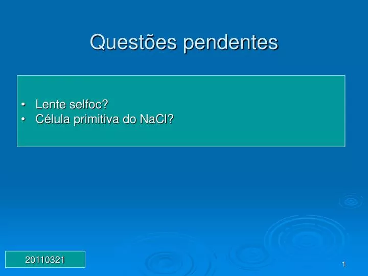
Questões pendentes. Lente selfoc? Célula primitiva do NaCl?. 20110321. Aula passada. Rede. Rede de Bravais. Rede cristalina = rede+base Empacotamento fechado Novas estruturas tipo buckyball Lei de Bragg. Índices de Miller. Fator de estrutura. Efetivamente como seria no NaCl?.

E N D
Questões pendentes • Lente selfoc? • Célula primitiva do NaCl? 20110321
Aula passada • Rede. Rede de Bravais. Rede cristalina = rede+base • Empacotamento fechado • Novas estruturas tipo buckyball • Lei de Bragg. Índices de Miller. Fator de estrutura.
Efetivamente como seria no NaCl? Íons da célula unitária: Na+ => ½ 0 0, 0 ½ 0, 0 0 ½ e ½ ½ ½ Cl- => 0 0 0, ½ 0 ½, ½ ½ 0, 0 ½ ½ Fhkl = ?
Tudo o anterior foi com relação a sistemas cristalinos • E sistemas não-cristalinos? i.e. amorfos? Imagens de R-X?
WILLIAM CONRAD ROENTGEN – Raio-X FIRST X-RAY - 1895 - ANNA BERTHA ROENTGEN'S HAND Art of Roentgen's X-ray apparatus for imaging hand
Novas técnicas de imagem de R-X. Physics Today Julho 2000
Aplicações de Raio-x Raio-X na área analítica Difração do pó SAXS/WAXS (Small Angle X ray Scattering + Wide Angle Xray Scattering) Cristalografia de moléculas e proteínas Reflectometria e análise de filmes finos Espectroscopia de Absorção de Raio-X Fluorescência de Raio-X dispersiva em comprimento de onda (WDX) Microdifração de Raio-X de Laue
Aplicações de Raio-x Raio-X na área de imagem – óptica • Microscopia de Raio-X • Laser de Raio-X
Podemos resumir que a óptica de raio-X pode.... Alem de microscopia e laser de raio-x
Não é imagem é apenas difração de material policristalino Difração de Raio-x de pó xstalino Como seria com material monocristalino?
SAXS/WAXS (Small Angle X ray Scattering + Wide Angle Xray Scattering) WAXS => grau de cristaliniddae SAXS => informação strutural de macromoléculas (5-25nm)
Imagem - Óptica de Raios-X • Difração? • Refletores? • Lentes? • Refração? • Polarização? • Laser de Raios-X?
Comparação entre luz e raios-X (Na3AlF6) (TiO2) (Na3AlF6)
Bent Laue crystal Point focusing A single bent multilayer or Laue crystal provides only focusing in one dimension, i.e. a line focus. Point focusing is achieved by combination of either two multilayers or a Laue crystal and a multilayer, the scattering planes of the combined elements being perpendicular. The synchrotron working group mainpage
Interferômetro de raios-x. • Realçamento por difração, variações na refração dos raios-x na amostra produzem contraste por causa da intensidade do feixe que é refletido pelo cristal analisador depende do ângulo de incidência do feixe em relação ao ângulo de Bragg. • Contraste de fase em linha
Mostrando interferometria de raio-x • Imagem CCD. O lado esquerdo mostra um dos feixes, o direiro mostra franjas de interferência.
Imagem feita por Ralf Menk. Elettra Synchrotron Light Source. Utilizando uma rede cristalina perfeita como um filtro angular muito sensível. O contraste aqui, origina-se em pequenas diferenças no ângulo de refração dos raios-x saindo da amostra. Características dos tecidos moles tais como o pêlo e os bigodes podem ser claramente vistas.).
Gafanhoto Radiografia convencional Radiografia por contraste de fase em linha
J. Anat. (2003) 202, p.463-470 Fig. 2 (a) Conventional synchrotron radiograph of the great toe that includes the distal portion of the first metatarsal (M), the sesamoid bones (S), the proximal phalanx (PP) and the distal phalanx (DP). The osteoporotic bones are evident as are the sclerotic vessels. (b) DE image of the same specimen as in (a) taken at +1 of the rocking curve. The major soft tissue structures that can be identified here, which are not visible in the above radiograph, include the two major tendons of the toe, the fat pad under the ball of the foot (which has been displaced distally somewhat) and the skin. The asterisk indicates the location of the muscles and tendons plantar to the first metatarsal bone. These cannot be delineated from one another in this image.
The Crab Nebula, the remnant of a supernova explosion in 1054 AD pictured below, is usually the most brilliant object in the hard x-ray sky. It shines so steadily that x-ray astronomers use a unit called "Crabs" to define how bright other sources are (the flux from the Crab Nebula is, by definition, 1 Crab). The Sun usually registers less than 0.01 Crabs, although sometimes a powerful solar flare will make the Sun shine brightly at hard x-ray wavelengths. Black hole binary systems like XTE J1550 can also erupt and briefly upstage the Crab Nebula, the "Old Reliable" of x-ray astronomy. Another black hole, Cygnus X-1, is a persistent hard x-ray source that is also sometimes brighter than the Crab.
Esquema básico http://www.warren.usyd.edu.au/bulletin/NO51/synchrotron_graphic_800.jpg
X-rays at Sample: Critical energy 28.9 keV Beam divergence 1.5 mrad (Horizontal)0.06 mrad (Vertical) Energy range Si 311 : 8.4 ∼ 72.5 keVSi 111 : 5.0 ∼ 37.5 keVSi 511 : 13.5 ∼ 113.3 keV Beam size 1 about 75 mm (H) × 5 mm (V) in the experimental hutch 1 located in the experimental hall (Si 311) Beam size 2 about 300 mm (H) × 20 mm (V) in the hutches located in the biomedical imaging center (Si 311) http://www.spring8.or.jp/wkg/BL20B2/instrument/lang-en/INS-0000000314/instrument_summary_view 2p radianos 360o 1rad 57,29o 1mrad 0,05729o 1mrad 3,438’
LNLS - Campinas Energia Nominal de Operação 1.37 GeV Energia de injeção 500 MeV Corrente do Feixe de elétrons (máximo) 250 mA Circunferência 93.2 m Diâmetro médio 29.7 m
Policapilaridade - Óptica de Raios-X http://www.xos.com/mfgpros.htm
X-ray Glass Capillary Optics A. Bjeoumikhov, S. Bjeoumikhova* IfG Institut für Gerätebau , 12489 Berlin, Germany * BAM, 12205 Berlin, Germany
Microscopia de raio-X Center for X-Ray Optics, Ernest O. Lawrence Laboratory Berkeley, CA