Download
1 / 22
350 likes | 479 Vues
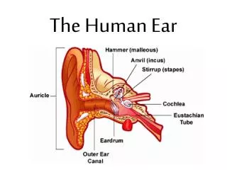
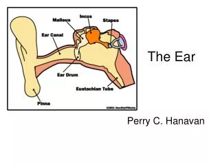
THE HUMAN EAR. SHERLI.K S PGT BIOLOGY KV No.II, KASARGOD. The ear performs two sensory functions: Hearing Maintenance of body balance . Anatomically, the ear can be divided into three major sections Outer Ear, Middle Ear and Inner Ear. STRUCTURE OF EAR. Structure of the Ear.

E N D
THE HUMAN EAR SHERLI.K S PGT BIOLOGY KV No.II, KASARGOD
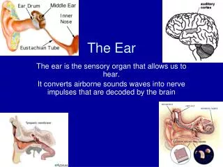
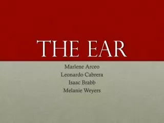
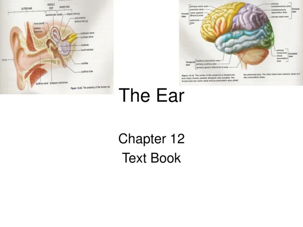
The ear performs two sensory functions: • Hearing • Maintenance of body balance. • Anatomically, the ear can be divided into three major sections • Outer Ear, • Middle Ear and • Inner Ear.
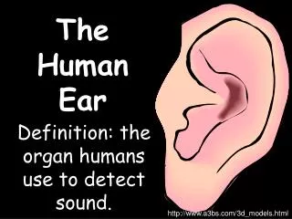
THE OUTER EAR • The Outer Ear consists of the Pinna and External Auditory Meatus (EAM). • The pinna collects the vibrations in the air which produce sound. • The EAM leads inwards and extends up to the Tympanic membrane( the ear drum). • Very fine hairs and sebaceous glands are present on the skin of the pinna. • Tympanic membrane is composed of connective tissues covered with skin outside and mucus membrane inside.
THE MIDDLE EAR • THE MIDDLE EAR -Consists of three ossicles called Malleus, Incus & Stapes. • Malleus is attached to Tympanic membrane and Stapes to The Oval Window of the Cochlea. • An Eustachian tube connects the middle ear cavity with the pharynx. • The Eustachian tube helps in equalising the pressures on either sides of the ear drum.
THE INNER EAR • The fluid filled inner ear called ‘LABYRINTH’ consists of two parts, the bony and the membranous labyrinths. • The bony labyrinth is a series of channels. Inside these channels lies the membranous labyrinth which is surrounded by a fluid called PERILYMPH. • The membranous labyrinth is filled with a fluid called ENDOLYMPH.
The coiled portion of the labyrinth is called cochlea. • The membranes constituting cochlea, the reissner’s and basilar, divide the surounding perilymph filled bony labyrinth into an upper scala vestibuli and a lower scala tympani . • The space within cochlea called scala media is filled with endolymph. • At the base of the cochlea, the scala vestibuli ends at the oval window, while the scala tympani terminates at the roundwindow which opens to the middle ear.
At the base of the cochlea, the scala vestibuli ends at the oval window, while the scala tympani terminates at the roundwindow which opens to the middle ear
The organ of corti is a structure located on the basilar membrane which contains hair cells that act as auditory receptors. • The hair cells are present in rows on the internal side of the organ of corti. • The basal end of the hair cell is in close contact with the afferent nerve fibres. • A large number of processes called stereo cilia are projected from the apical part of each hair cell. • Above the rows of the hair cells is a thin elastic membrane called tectorial membrane
Vestibular apparatus Vestibular apparatus, located above the cochlea. The vestibular apparatus is composed of three semi-circular canals and the otolith organ consisting of the saccule and utricle. Each semi-circular canal lies in a different plane at right angles to each other.
The membranous canals are suspended in the perilymph of the bony canals. The base of canals is swollen and is called ampulla, which contains a projecting ridge called crista ampullaris which has hair cells. The saccule and utricle contain a projecting ridge called macula. The crista and macula are the specific receptors of thevestibular apparatus responsible for maintenance of balance of the body and posture.
REVIEW QUESTIONS • 1.Three major sections of ear are ---------, --------and---------. • 2.---------connects the middle ear cavity with the pharynx. • 3The membranous labyrinth is filled with a fluid called --------- • 4. ------------ part of our ear helps in maintaining the body balance and posture. • 5. The coiled portion of the labyrinth is called --------