Download
1 / 45
600 likes | 1.11k Vues
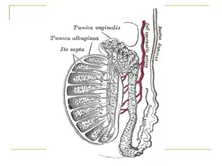
Testicular diseases. Epididymitis And ORCHITIS : Inflammatory conditions are generally more common in the epididymis than in the testis However, some infections,notably syphilis,may begin in the testis with secondary involvement of the epididymis. Normal Anatomy.

E N D
Testicular diseases Epididymitis And ORCHITIS: Inflammatory conditions are generally more common in the epididymis than in the testis However, some infections,notably syphilis,may begin in the testis with secondary involvement of the epididymis
Epididymitis And ORCHITIS 1.Non specific epididymitis and Orchitis: Epididymitis and possible subsequent orchitis are commonly related to infections in the urinarytract(cystitis,urethritis,genitoprostatitis. These infections reach the epididymis/testis through either the vas deference or the lymphatics of the spermatic cord.
Epididymitis And ORCHITIS Epididymitis CAUSES: Varies with age; Children: uncommon, usually associated with a congenital genitourinary abnormality and infection with Gram –ve rods. In sexually active men younger than age 35 years-----Chlamydia trachomatis and Neisseria
Epididymitis And ORCHITIS • Older than 35 ---------E.Coli and Pseudomonos. • Microscopic findings: • Bacterial invasion Non specific acute inflammation characterized by congestion,edema and infiltration by neutrophils,macrophages and lymphocytes. • Initially involves the interstitial connective tissue later involves tubules may progress to frank abscess.
Epididymitis And ORCHITIS often followed by fibrous scarring. Leydig cells are not usually destroyed.
2.Granulomatous(Autoimmune) Orchitis Usually middle –aged men------unilateral testicular mass. Usually moderately tender but sometimes may present as painless testicular mass; mimicking a testicular tumor. Microscopically: granulomas resticted within the spermatic tubules. Although an autoimmune basis is suspected,the cause of these lesions remain unknown.
May be a response to acid-fast products of disintegrated sperm, post-infectious, or due to trauma or sarcoidosis
3.Specific Inflammations: Gonorrhea: Extension of infection from the posterior urethra to the prostate to the seminal vesicles and then to the epididymis is the usual course of a neglected gonococcal infection. • Can lead to frank abscess may spread to testis and can produce a suppurative orchitis.
Specific Inflammations: Tuberculosis: • Almost invariably begins in the epididymis and may spread to the testis. • In many of these cases ,there is associated tuberculous prostatitis and seminal vesiculitis and it is believed that epididymitis usually represents a secondary spread from these other involvements of the genital tract
Specific Inflammations: Microscopy: Caseating Granulomatous inflammation.
Specific Inflammations: • Syphilis: • Affected in both acquired and congenital syphilis. • Almost invariably, the testis is involved first by the infection. • Morphology: Gummas production or • diffuse interstitial inflammation characterized by edema and lymphocytic and plasma cell infiltration.
Specific Inflammations: Obliterative endarteritis with perivascular cuffing of lymphocytes and plasma cells.
Testicular Tumors • Complex mixture of anatomic types • 95% of them originate from germ cells • Most of gem cell tumors are highly aggressive cancers • Capable of wide ,extensive dissemination • Current therapy ,most of them can be cured
Testicular TumorsGerm cell types • World wide increase in the incidence of of these tumors • At age 15-30 these are the most common tumor of men • More common in whites than blacks.
Testicular Tumors Classification • Germ cell tumors : -Seminoma -Spermatocytic seminoma -Embryonal carcinoma -Yolk sac (endodermal Sinus) tumor -Choriocarcinoma -Teratoma
Testicular Tumors Classification • Sex Cord Tumors -Leydig cell tumor -Sertoli cell tumor
Testicular Tumors Pathogenesis • Several predisposing factors: -Cryptorchidism :10% of testicular tumors -Testicular dysgenesis -Genetic factors
Seminoma • The most common type of germ cell tumors 50% • Almost never occur in infants • Peak incidence in thirties • Identical one occurs in the ovary(Dysgerminoma)
Seminoma , Morphology • Refers to the classic or typical seminoma • Bulky masses • Homogenous ,gray-white ,lobulated cut surface • Usually no necrosis or hemorrhage • In 50% ,the entire testis is involved • Occasionally extends to the epididymis,spermatic cord,or scrotal sac
Seminoma , Morphology • Microscopically ,sheets of uniform cells • Lobules,separated by delicate fibrous septa • Cells are large ,round ,has distinct cell membrane • Large nucleus with prominent nucleolei • Positive for PLAP
Spermatocytic Seminoma • Distinctive tumor ,clinically and histologically • 1-2 % of testicular tumors • Over age 65 • Slow growing tumor ,rarely metastasise • Prognosis is excellent
Embryonal Carcinoma • 20 to 30 year age group • More aggressive than seminomas • Smaller than seminoma • Foci of necrosis and hemorrhage • Cells grow in alveolar or tubular pattern ,papillary convolutions • Could be present with other neoplasms45%
Yolk Sac Tumor • Infantile embryonal carcinoma • Endodermal sinus tumor • The most common tumor in infant and children up to 3 years of age • Has a very good prognosis • Non encapsulated ,homogenous ,mucinous appearance
Yolk Sac Tumor • Microscopically ,structures resemble endodermal sinuses • Schiller-Duval bodies • Hyaline –pink globules • AFP positive
Choriocarcinoma • Highly malignant tumor • Cytotrophoblastic and syncytiotroblastic cells • Small lesions • HCG positive
Teratoma • Various cellular or organoid components • Any age ,infancy to adult life • Mature forms are common in infants and children • Second to yolk sac tumor in this age group • Adult forms are rare • As a component with other type 45%
Teratoma • Usually large 5 -10 cm • Heterogenous appearance • Hemorrhage and necrosis indicate embryonal component • Composedof heterogenous collection of cells or organoid structures • Neural tissue ,cartilage ,squamous epithelium ,glandular components….
teratoma • Germ cell tumors could arise from teratoma • In children ,mature teratomas behave benign • In post pubertal male ,all teratomas regarded malignant ,and capable of metastasis ,regardless of of whether the elements are mature or not
Testicular tumorsClinical Features • Biopsy of a testicular tumor is associated with a risk of tumor spillage • The standard manaagment of solid tumors is radical orchiectomy • Lymphatic spread is common • Retroperitoneal and para-aortic nodes are first to be involved • Hematogenous spread to Lung, liver, Brain, and bones.