Download
1 / 2
20 likes | 128 Vues
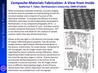
Composite Materials Fabrication: A View from Inside Katherine T. Faber , Northwestern University, DMR 0710630.

E N D
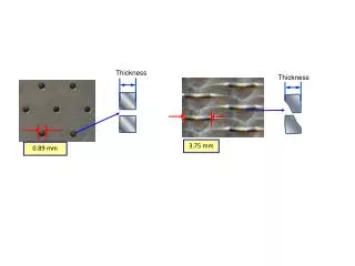
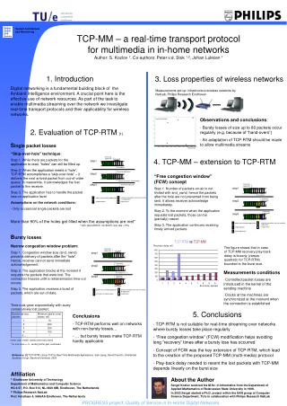
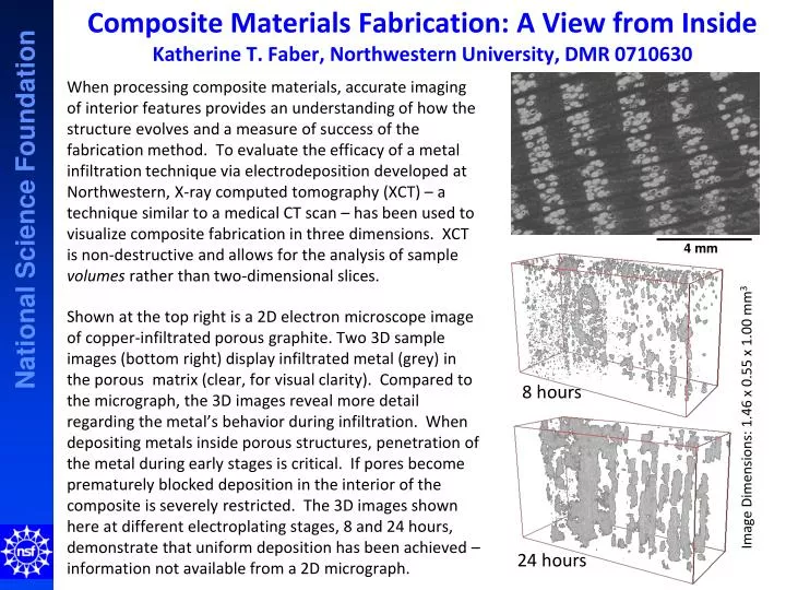
Composite Materials Fabrication: A View from InsideKatherine T. Faber, Northwestern University, DMR 0710630 When processing composite materials, accurate imaging of interior features provides an understanding of how the structure evolves and a measure of success of the fabrication method. To evaluate the efficacy of a metal infiltration technique via electrodeposition developed at Northwestern, X-ray computed tomography (XCT) – a technique similar to a medical CT scan – has been used to visualize composite fabrication in three dimensions. XCT is non-destructive and allows for the analysis of sample volumes rather than two-dimensional slices. Shown at the top right is a 2D electron microscope image of copper-infiltrated porous graphite. Two 3D sample images (bottom right) display infiltrated metal (grey) in the porous matrix (clear, for visual clarity). Compared to the micrograph, the 3D images reveal more detail regarding the metal’s behavior during infiltration. When depositing metals inside porous structures, penetration of the metal during early stages is critical. If pores become prematurely blocked deposition in the interior of the composite is severely restricted. The 3D images shown here at different electroplating stages, 8 and 24 hours, demonstrate that uniform deposition has been achieved – information not available from a 2D micrograph. 8 hours Image Dimensions: 1.46 x 0.55 x 1.00 mm3 4 mm 24 hours
Materials World Networking in SevilleKatherine T. Faber, Northwestern University, DMR-0710630 An integral part of the Materials World Network (MWN) Program is the technical exchange of students among participating institutions. While graduate student Matthew Johnson paid a research visit to the University of Seville, he participated in their annual day-long seminar hosted by MWN collaborators. The seminar, entitled “Ceramic Engineering for Extreme Environments,” (program shown at right) featured talks from Spanish researchers covering materials and characterization of solid-liquid interfaces to fiber composites. Matthew presented his work on thermal analysis and 3D imaging of wood-derived graphite-carbon composites. Sharing these results with some of the Spanish researchers who pioneered work on the processing of wood-derived ceramic materials proved vital for continued collaboration.