Download
1 / 33
350 likes | 1.08k Vues
Atherosclerosis. Anca Ba cârea , Alexandru Schiopu. Atherosclerosis.

E N D
Atherosclerosis Anca Bacârea, Alexandru Schiopu
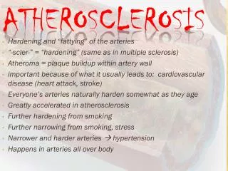
Atherosclerosis • The term atherosclerosis, which comes from the Greek words atheros (meaning “gruel” or “paste”) and sclerosis (meaning “hardness”), denotes the formation of fibrofatty lesions in the intimal lining of the large and medium-size arteries such as the aorta and its branches, the coronary arteries, and the large vessels that supply the brain. • Atherosclerosis contributes to more mortality and more serious morbidity than any other disorder in the western world.
Risk Factors • The cause or causes of atherosclerosis have not been determined with certainty. • Epidemiologic studies have identified predisposing risk factors: • Unchangeable risk factors • Age • Male gender • Men are at grater risk than are premenopausal women, because of the protective effects of natural estrogens. • Family history of premature coronary heart disease • Several genetically determined alterations in lipoprotein and cholesterol metabolism have been identified.
Risk Factors • Changeable risk factors – life style risk factors: • Hyperlipidemia • The presence of hyperlipidemia is the strongest risk factor for atherosclerosis in persons younger than 45 years of age. • Both primary and secondary hyperlipidemia increase the risk. • Cigarette smoking • Hypertension • High blood pressure produces mechanical stress on the vessel endothelium. • It is a major risk factor for atherosclerosis in all age groups and may be as important or more important than hypercholesterolemia after the age of 45 years. • Diabetes mellitus • Diabetes elevates blood lipid levels and otherwise increases the risk of atherosclerosis. • Insufficient physical activity • A stressful lifestyle • Obesity
Risk Factors • There are a number of other less well-established risk factors for atherosclerosis, including: • High serum homocysteine levels • Homocysteine is derived from the metabolism of dietary methionine • Homocysteine inhibits elements of the anticoagulant cascade and is associated with endothelial damage. • Elevated serum C-reactive protein • It may increase the likelihood of thrombus formation; • Inflammation marker; • Infectious agents • The presence of some organisms (Chlamydia pneumoniae, herpesvirus hominis, cytomegalovirus) in atheromatous lesions has been demonstrated by immunocytochemistry, but no cause-and-effect relationship has been established. • The organisms may play a role in atherosclerotic development by initiating and enhancing the inflammatory response.
Pathology and pathogenesis • The lesions associated with atherosclerosis are of three types: • The fatty streak • The fibrous atheromatous plaque • Complicatedlesion • The latter two are responsible for the clinicallysignificant manifestations of the disease.
Pathology and pathogenesis • Fatty streaks are thin, flat yellow intimal discolorations thatprogressively enlarge by becoming thicker and slightly elevatedas they grow in length. • They consistof macrophages and smooth muscle cells that have become distendedwith lipid to form foam cells. • These occurs regardless of geographic setting, gender, or race. • They increase in number until about age 20 years, and then they remain static or regress. • There is controversy about whether fatty streaks, in and of themselves, are precursors of atherosclerotic lesions.
Pathology and pathogenesis • The fibrous atheromatous plaque is the basic lesion of clinicalatherosclerosis. • It is characterized by the accumulation of intracellularand extracellular lipids, proliferation of vascularsmooth muscle cells, and formation of scar tissue. • The lesionsbegin as a elevated thickening of the vesselintima with a core of extracellular lipid (mainly cholesterol,which usually is complexed to proteins) covered by a fibrouscap of connective tissue and smooth muscle. • As thelesions increase in size, they encroach on the lumen of the arteryand eventually may occlude the vessel or predispose to thrombusformation, causing a reduction of blood flow.
Pathology and pathogenesis • The more advanced complicated lesions are characterized by • Hemorrhage • Ulceration • Scar tissue deposits • Thrombosisis the most important complication of atherosclerosis. • It is caused by slowing and turbulence of blood flow in the regionof the plaque and ulceration of the plaque.
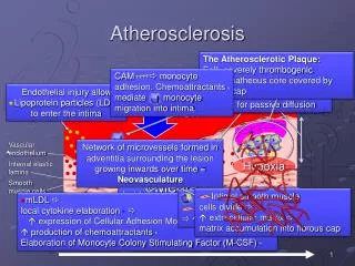
Mechanisms • There is increasing evidence that atherosclerosis is at leastpartially the result of: • (1) endothelial injury with leukocyte(lymphocyte and monocyte) adhesion and platelet adherence • (2) smooth muscle cell emigration and proliferation • (3) lipid engulfmentof activated macrophages • (4) subsequent developmentof an atherosclerotic plaque with lipid core
Mechanisms • One hypothesis of plaque formation suggests that injury to the endothelial vessel layer is the initiating factor in the development of atherosclerosis. • Possible injurious agents are: • Products associated with smoking; • Immune mechanisms; • Mechanical stress, such as that associated with hypertension. • Hyperlipidemia, particularly LDL with its high cholesterol content, is also believed to play an active role in the pathogenesis of the atherosclerotic lesion.
LDL - cholesterol • The LDLis removed from the circulation by either LDL receptors or byscavenger cells such as monocytes or macrophages. • Approximately70% of LDL is removed by way of the LDL receptordependentpathway.Although LDL receptors are widely distributed,approximately 75% are located on hepatocytes; thusthe liver plays an extremely important role in LDL metabolism. • Tissues with LDL receptors can control their cholesterol intakeby adding or removing LDL receptors. • The scavenger cells, such as the monocytes and macrophages,have receptors that bind LDL that has been oxidized orchemically modified. • The amount of LDL that is removed bythe “scavenger pathway” is directly related to the plasma cholesterollevel. When there is a decrease in LDL receptors orwhen LDL levels exceed receptor availability, the amount ofLDL that is removed by scavenger cells is greatly increased. • Theuptake of LDL by macrophages in the arterial wall can result inthe accumulation of insoluble cholesterol esters, the formationof foam cells, and the development of atherosclerosis.
Mechanisms • One of the earliest responses to elevatedcholesterol levels is the attachment of monocytes to theendothelium. • The monocytes emigratethrough the cell-to-cell attachments of the endothelial layerinto the subendothelial spaces, where they are transformedinto macrophages. • Activated macrophages release free radicalsthat oxidize LDL. • Oxidized LDL is not recognized at the cell receptor level and so, it can not be internalized and it longer remains into the blood stream. • Oxidized LDL is toxic to the endothelium,causing endothelial loss and exposure of the subendothelialtissue to blood components: • It has chemotactic effect on lymphocytes and monocytes; • It has chemotactic effect on smoothmuscle cells from the arterial media and stimulates production of MG-CSF, cytokines, adhesion molecules in the endothelium; • It inhibits endothelium derived releasing factor (EDRF), favoring vasospasm; • It stimulates specific immune system (production of antibodies against oxidized LDL).
Mechanisms • Endothelial disruption leads to platelet adhesionand aggregation and fibrin deposition. • Platelets and activatedmacrophages release various factors that are thought to promotegrowth factors that modulate the proliferation of smoothmuscle cells and deposition of extracellular matrix in the lesions: elastin, collagen, proteoglycans. • Activated macrophages also ingest oxidized LDL to becomefoam cells, which are present in all stages of atheroscleroticplaque formation. • Lipids released from necrotic foamcells accumulate to form the lipid core of unstable plaques. • Connective tissue synthesis determinates stiffness, calcium fixation and further ulceration of atheromatous plaque.
Glycosylation and atherosclerosis • Glycosylation is a process that affects lipoproteins, circulating proteins and proteins component of the arterial wall. • Effects: • Glycated LDL stimulates platelet aggregation and forms covalent bounds with the proteins of the arterial wall. • Glycated HDL blocks cholesterol efflux from the cells. • Collagen glycosylation increases arterial wall stiffness, activates macrophages and stimulates lipoprotein adherence. • Glycosylated proteins form circulating antigens which generates antibody and circulating immune complexes that will lead to other arterial lesions.
Modern theory of atherosclerosis • Multifactor theory: • Structural and functional injury of vascular endothelium; • Response to injury of immune cells and smooth muscle cells; • The role of lipoproteins in initiation and progression of lesions; • The role of growth factors and cytokines; • The role of repeated thrombosis in lesions progression.
Mechanisms • As a result of all presented above atherosclerosis can be defined as vicious inflammatory process.
Clinical Manifestations • The clinical manifestations of atherosclerosis depend on the vessels involved and the extent of vessel obstruction. • Atherosclerotic lesions produce their effects through: • narrowing of the vessel and production of ischemia; • sudden vessel obstruction caused by plaque hemorrhage or rupture; • thrombosis and formation of emboli resulting from damage to the vessel endothelium; • In larger vessels such as the aorta, the important complications are those of thrombus formation and weakening of the vessel wall. • In medium-size arteries such as the coronary and cerebral arteries, ischemia and infarction caused by vessel occlusion are more common. • Although atherosclerosis can affect any organ or tissue, the arteries supplying the heart, brain, kidneys, lower extremities, and small intestine are most frequently involved.
Coronary heart disease • The term coronary heart disease (CHD) describes heart diseasecaused by impaired coronary blood flow. • In most cases, it is caused by atherosclerosis. • Diseases of the coronary arteriescan cause: • Angina • Myocardial infarction or heart attack • Cardiac dysrhythmias • Conduction defects • Heart failure • Suddendeath
Coronary circulation • There are two main coronary arteries, the left and the right, which arise from the coronary sinus just above the aortic valve. • Although there are no connections between the large coronary arteries, there are anastomotic channels that join the small arteries. • The primary factor responsible for perfusion of the coronary arteries is the aortic blood pressure. • Changes in aortic pressure produce parallel changes in coronary blood flow. • The contracting heart muscle influences its own blood supply by compressing the intramyocardial and subendocardial blood vessels. As a result, blood flow through the subendocardial vessels occurs mainly during diastole. • Thus, there is increased risk of subendocardial ischemia when a rapid heart rate decreases the time spent in diastole, and when an elevation in diastolic intraventricular pressure is sufficient to compress the vessels in the subendocardial plexus.
Coronary circulation • Blood flow usually is regulated by the need of the cardiac muscle for oxygen. • Even under normal resting conditions, the heart extracts and uses 60% to 80% of oxygen in blood flowing through the coronary arteries, compared with the 25% to 30% extracted by skeletal muscle. • Because there is little oxygen reserve in the blood, myocardial ischemia develops when the coronary arteries are unable to dilate and increase blood flow during periods of increased activity or stress. • Heart muscle relies primarily on fatty acids and aerobic metabolism to meet its energy needs. Although the heart can engage in anaerobic metabolism, this process relies on the continuous delivery of glucose and results in the formation of large amounts of lactic acid.
Pathogenesis of coronary heart disease (CHD) • Atherosclerosis is by far the mostcommon cause of CHD, and atherosclerotic plaque disruptionthe most frequent cause of myocardial infarction and suddendeath. • More than 90% of persons with CHD have coronary atherosclerosis. • Most, if not all, have one or more lesions causingat least 75% reduction in cross-sectional area, the point atwhich augmented blood flow provided by compensatory vasodilationno longer is able to assure even moderate increasesin metabolic demand. • There are two types of atherosclerotic lesions: • the fixed orstable plaque, which obstructs blood flow • commonly implicated in chronic ischemic heart disease: stable angina, variant or vasospastic angina, and silent myocardial ischemia; • the unstable orvulnerable plaque, which can rupture and cause platelet adhesionand thrombus formation • commonly implicated in unstable anginaand myocardial infarction.
Pathogenesis of coronary heart disease (CHD) • Plaque disruption may occur with or without thrombosis. • Platelets play a major role in linking plaque disruption to acute CHD. • As a part of the response to plaque disruption, platelets aggregate and release substances that further propagate platelet aggregation, vasoconstriction, and thrombus formation. • Because of the role that platelets play in the pathogenesis of CHD, antiplatelet drugs (e.g., low-dose aspirin) are frequently used for preventing heart attack.
Myocardial infarction • Acute myocardial infarction (AMI), also known as a heartattack, is characterized by the ischemic death of myocardialtissue associated with atherosclerotic disease of the coronaryarteries. • Diagnosis: • 1. Pain • The pain typically is severe and crushing, often described as being constricting, suffocating. It usually is substernal, radiating to the left arm, neck, or jaw, although it may be experienced in other areas of the chest. • Gastrointestinal complaints are common. There may be a sensation of epigastric distress; nausea and vomiting may occur. • 2. ECG • Elevation of the ST segment usually indicates acutemyocardial injury. • When the ST segment is elevated withoutassociated Q waves, it is called a non–Q-wave infarction. Anon–Q-wave infarction usually represents a small infarct thatmay evolve into a larger infarct. • 3. Enzymes
Enzymes • Myoglobin is an oxygen-carrying protein, similar to hemoglobin,that is normally present in cardiac and skeletal muscle. Itis a small molecule that is released quickly from infarcted myocardialtissue and becomes elevated within 1 hour after myocardialcell death, with peak levels reached within 4 to 8 hours. It rapidly eliminates through urine (low molecular weight). Because myoglobin is present in both cardiac and skeletalmuscle, it is not cardiac specific. • Creatine kinase (CK), formerly called creatinine phosphokinase,is an intracellular enzyme found in muscle cells. Muscles, includingcardiac muscle, use ATP astheir energy source. Creatine, which serves as a storage formof energy in muscle, uses CK to convert ADP to ATP. CK exceedsnormal range within 4 to 8 hours of myocardial injuryand declines to normal within 2 to 3 days. There are threeisoenzymes of CK, with the MB isoenzyme (CK-MB) beinghighly specific for injury to myocardial tissue.
Enzymes • The troponin complex consists of three subunits (i.e., troponinC, troponin I, and troponin T) that regulate calcium-mediatedcontractile process in striated muscle. These subunits are releasedduring myocardial infarction. Cardiac muscle forms ofboth troponin T and troponin I are used in diagnosis of myocardialinfarction. Troponin I (and troponin T; not shown) risesmore slowly than myoglobin and may be useful for diagnosisof infarction, even up to 3 to 4 days after the event. It isthought that cardiac troponin assays are more capable ofdetecting episodes of myocardial infarction in which celldamage is below that detected by CK-MB level.
Effects of AMI • The principal biochemical consequence of AMI is the conversionfrom aerobic to anaerobic metabolism with inadequateproduction of energy to sustain normal myocardial function. • The ischemic area ceases to function within amatter of minutes, and irreversible myocardial cell damage occursafter 20 to 40 minutes of severe ischemia. • The term reperfusion refers to re-establishment of blood flowthrough use of thrombolytic therapy or revascularization procedures. • Early reperfusion (within 15 to 20 minutes) after onsetof ischemia can prevent necrosis. • Reperfusion after a longer intervalcan salvage some of the myocardial cells that would havedied because of longer periods of ischemia.
Peripheral arterial disease (PAD) • PAD refers to the obstruction of large arteries not within the coronary, aortic arch vasculature, or brain. • It can result from atherosclerosis, inflammatory processes leading to stenosis, an embolism, or thrombus formation. • It causes either acute or chronic ischemia (lack of blood supply). • Often PAD is a term used to refer to atherosclerotic blockages found in the lower extremity. • Risk factors contributing to PAD are the same as those for atherosclerosis. • Risk of PAD also increases in individuals who are over the age of 50, male, obese, or with a family history of vascular disease, heart attack, or stroke.
Peripheral arterial disease (PAD) • About 20% of patients with mild PAD may be asymptomatic; • Symptoms include: • Claudication - pain, weakness, numbness, or cramping in muscles due to decreased blood flow; • Sores, wounds, or ulcers that heal slowly or not at all ; • Noticeable change in color (blueness or paleness) or temperature (coolness) when compared to the other limb ; • Diminished hair and nail growth on affected limb and digits.