Download
1 / 121
1.32k likes | 2.1k Vues
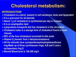
Cholesterol and Steroid Metabolism . UNIT III: Lipid Metabolism. I. Overview . Cholesterol, the characteristic steroid alcohol of animal tissues, performs a number of essential functions in the body.

E N D
Cholesterol and Steroid Metabolism UNIT III: Lipid Metabolism
I. Overview • Cholesterol, the characteristic steroid alcohol of animal tissues, performs a number of essential functions in the body. • For example, cholesterol is a structural component of all cell membranes, modulating their fluidity, and, in specialized tissues, cholesterol is a precursor of bile acids, steroid hormones, and vitamin D. • It is therefore of critical importance that the cells of the body be assured an appropriate supply of cholesterol. • To meet this need, a complex series of transport, biosynthetic, and regulatory mechanisms has evolved. • The liver plays a central role in the regulation of the body's cholesterol homeostasis.
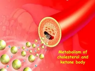
For example, cholesterol enters the liver's cholesterol pool from a number of sources including dietary cholesterol, as well as cholesterol synthesized de novo by extrahepatic tissues and by the liver itself. • Cholesterol is eliminated from the liver as unmodified cholesterol in the bile, or it can be converted to bile salts that are secreted into the intestinal lumen. • It can also serve as a component of plasma lipoproteins sent to the peripheral tissues. • In humans, the balance between cholesterol influx and efflux is not precise, resulting in a gradual deposition of cholesterol in the tissues, particularly in the endothelial linings of blood vessels. • This is a potentially life-threatening occurrence when the lipid deposition leads to plaque formation, causing the narrowing of blood vessels (atherosclerosis) and increased risk of coronary artery disease.
The major sources of liver cholesterol and the routes by which cholesterol leaves the liver
II. Structure of Cholesterol • Cholesterol is a very hydrophobic compound. It consists of four fused hydrocarbon rings (A, B, C, and D, called the “steroid nucleus”), and it has an eight-carbon, branched hydrocarbon chain attached to C-17 of the D ring. • Ring A has a hydroxyl group at C-3, and ring B has a double bond between C-5 and C-6 (Figure 18.2).
Sterols • Steroids with 8–10 carbon atoms in the side chain at C-17 and a hydroxyl group at C-3 are classified as sterols. • Cholesterol is the major sterol in animal tissues. • [Note: Plant sterols, such as β-sitosterol are poorly absorbed by humans. After entering the enterocytes, they are actively transported back into the intestinal lumen. Because some cholesterol is transported as well, plant sterols appear to block the absorption of dietary cholesterol. This has led to clinically useful dietary treatment of hypercholesteremia. Daily ingestion of plant steroid esters (in the form of commercially available trans fatty acid–free margarine) is one of a number of dietary strategies leading to the reduction of plasma cholesterol levels]
B. Cholesteryl esters • Most plasma cholesterol is in an esterified form (with a fatty acid attached at C-3), which makes the structure even more hydrophobic than free cholesterol. • Cholesteryl esters are not found in membranes, and are normally present only in low levels in most cells. • Because of their hydrophobicity, cholesterol and its esters must be transported in association with protein as a component of a lipoprotein particle or be solubilized by phospholipids and bile salts in the bile.
III. Synthesis of Cholesterol • Cholesterol is synthesized by virtually all tissues in humans, although liver, intestine, adrenal cortex, and reproductive tissues, including ovaries, testes, and placenta, make the largest contributions to the body's cholesterol pool. • As with fatty acids, all the carbon atoms in cholesterol are provided by acetate, and NADPH provides the reducing equivalents. • The pathway is endergonic, being driven by hydrolysis of the high-energy thioester bond of acetyl coenzyme A (CoA) and the terminal phosphate bond of adenosine triphosphate (ATP).
Synthesis occurs in the cytoplasm, with enzymes in both the cytosol and the membrane of the endoplasmic reticulum. • The pathway is responsive to changes in cholesterol concentration, and regulatory mechanisms exist to balance the rate of cholesterol synthesis within the body against the rate of cholesterol excretion. • An imbalance in this regulation can lead to an elevation in circulating levels of plasma cholesterol, with the potential for coronary artery disease.
Synthesis of 3-hydroxy-3-methylglutaryl (HMG) CoA • The first two reactions in the cholesterol synthetic pathway are similar to those in the pathway that produces ketone bodies. • They result in the production of HMG CoA. • First, two acetyl CoA molecules condense to form acetoacetyl CoA. • Next, a third molecule of acetyl CoA is added, producing HMG CoA, a six-carbon compound. • [Note: Liver parenchymal cells contain two isoenzymes of HMG CoA synthase. The cytosolic enzyme participates in cholesterol synthesis, whereas the mitochondrial enzyme functions in the pathway for ketone body synthesis.]
B. Synthesis of mevalonic acid (mevalonate) • The next step, the reduction of HMG CoA to mevalonic acid, is catalyzed by HMG CoA reductase, and is the rate-limiting and key regulated step in cholesterol synthesis. • It occurs in the cytosol, uses two molecules of NADPH as the reducing agent, and releases CoA, making the reaction irreversible. • [Note: HMG CoA reductase is an intrinsic membrane protein of the endoplasmic reticulum (ER), with the enzyme's catalytic domain projecting into the cytosol.
C. Synthesis of cholesterol • The reactions and enzymes involved in the synthesis of cholesterol from mevalonate are illustrated in Figure 18.5. • [Note: The numbers shown in brackets below correspond to numbered reactions shown in this figure.]
[1] Mevalonic acid is converted to 5-pyrophosphomevalonate in two steps, each of which transfers a phosphate group from ATP. [2] A five-carbon isoprene unit—isopentenyl pyrophosphate (IPP)—is formed by the decarboxylation of 5-pyrophosphomevalonate. The reaction requires ATP. • [Note: IPP is the precursor of a family of molecules with diverse functions, the isoprenoids. Cholesterol is a sterol isoprenoid. Nonsterol isoprenoids include dolichol and ubiquinone.]
[3] IPP is isomerized to 3,3-dimethylallyl pyrophosphate (DPP). [4] IPP and DPP condense to form ten-carbon geranyl pyrophosphate (GPP). [5] A second molecule of IPP then condenses with GPP to form 15-carbon farnesyl pyrophosphate (FPP). • [Note: Covalent attachment of farnesyl to proteins, a process known as “prenylation,” is one mechanism for anchoring proteins to plasma membranes.] [6] Two molecules of FPP combine, releasing pyrophosphate, and are reduced, forming the 30-carbon compound squalene. • [Note: Squalene is formed from six isoprenoid units. Because three ATP are hydrolyzed per mevalonic acid residue converted to IPP, a total of 18 ATP are required to make the polyisoprenoid squalene.]
[7] Squalene is converted to the sterol lanosterol by a sequence of reactions that use molecular oxygen and NADPH. The hydroxylation of squalene triggers the cyclization of the structure to lanosterol. [8] The conversion of lanosterol to cholesterol is a multistep process, resulting in the shortening of the carbon chain from 30 to 27 carbons, removal of the two methyl groups at C-4, migration of the double bond from C-8 to C-5, and reduction of the double bond between C-24 and C-25. • [Note: This pathway includes more than 19 different enzymatic reactions. Smith-Lemli-Opitz syndrome (SLOS), a relatively common autosomal recessive disorder of cholesterol biosynthesis, is caused by a partial deficiency in 7-dehydrocholesterol-7-reductase—an enzyme involved in the migration of the double bond. SLOS is one of several multisystem, embryonic malformation syndromes associated with impaired cholesterol synthesis.]
D. Regulation of cholesterol synthesis • HMG CoA reductase, the rate-limiting enzyme, is the major control point for cholesterol biosynthesis, and is subject to different kinds of metabolic control. • Sterol-dependent regulation of gene expression: • Expression of the HMG CoA reductase gene is controlled by the transcription factor, SREBP (sterol regulatory element–binding protein) that binds DNA at the cis-acting sterol regulatory element (SRE) of the reductase gene. • SREBP is an integral protein of the ER membrane, and associates with a second ER membrane protein, SCAP (SREBP cleavage–activating protein).
Figure 18.6 Regulation of HMG CoA reductase. SRE = sterol regulatory element; SREBP = sterol regulatory element binding protein; SCAP = SREBP cleavage-activating protein
2. Sterol-accelerated enzyme degradation: • The reductase itself is an integral protein of the ER membrane. When sterol levels in the cell are high, the reductase binds to insig proteins. This binding leads to ubiquitination and proteasomal degradation of the reductase. 3. Sterol-independent phosphorylation/dephosphorylation: • HMG CoA reductase activity is controlled covalently through the actions of adenosine monophosphate (AMP)–activated protein kinase (AMPK), and a phosphoprotein phosphatase. • The phosphorylated form of the enzyme is inactive, whereas the dephosphorylated form is active.
[Note: AMPK is activated by AMP, so cholesterol synthesis, like fatty acid synthesis, is decreased when ATP availability is decreased.] 4. Hormonal regulation: • The amount (and, therefore, the activity) of HMG CoA reductase is controlled hormonally. • An increase in insulin favors up-regulation of the expression of the HMG CoA reductase gene. Glucagon has the opposite effect.
5. Inhibition by drugs: • The statin drugs (atorvastatin, fluvastatin, lovastatin, pravastatin, rosuvastatin, and simvastatin) are structural analogs of HMG CoA, and are (or are metabolized to) reversible, competitive inhibitors of HMG CoA reductase. • They are used to decrease plasma cholesterol levels in patients with hypercholesterolemia
Figure 18.7 Structural similarity of HMG and simvastatin, a clinically useful cholesterol-lowering drug of the “statin” family.
IV. Degradation of Cholesterol • The ring structure of cholesterol cannot be metabolized to CO2 and H2O in humans. • Rather, the intact sterol nucleus is eliminated from the body by conversion to bile acids and bile salts, which are excreted in the feces, and by secretion of cholesterol into the bile, which transports it to the intestine for elimination. • Some of the cholesterol in the intestine is modified by bacteria before excretion. The primary compounds made are the isomers coprostanol and cholestanol, which are reduced derivatives of cholesterol. • Together with cholesterol, these compounds make up the bulk of neutral fecal sterols.
V. Bile Acids and Bile Salts • Bile consists of a watery mixture of organic and inorganic compounds. • Phosphatidylcholine (lecithin) and bile salts (conjugated bile acids) are quantitatively the most important organic components of bile. • Bile can either pass directly from the liver where it is synthesized into the duodenum through the common bile duct, or be stored in the gallbladder when not immediately needed for digestion.
A. Structure of the bile acids • The bile acids contain 24 carbons, with two or three hydroxyl groups and a side chain that terminates in a carboxyl group. • The carboxyl group has a pKa of about six and, therefore, is not fully ionized at physiologic pH—hence, the term “bile acid.” • The bile acids are amphipathic in that the hydroxyl groups are α in orientation (they lie “below” the plane of the rings) and the methyl groups are β (they lie “above” the plane of the rings). • Therefore, the molecules have both a polar and a nonpolar face, and can act as emulsifying agents in the intestine, helping prepare dietary triacylglycerol and other complex lipids for degradation by pancreatic digestive enzymes.
B. Synthesis of bile acids • Bile acids are synthesized in the liver by a multistep, multiorganelle pathway in which hydroxyl groups are inserted at specific positions on the steroid structure, the double bond of the cholesterol B ring is reduced, and the hydrocarbon chain is shortened by three carbons, introducing a carboxyl group at the end of the chain. • The most common resulting compounds, cholic acid (a triol) and chenodeoxycholic acid (a diol), are called “primary” bile acids. • [Note: The rate-limiting step in bile acid synthesis is the introduction of a hydroxyl group at carbon 7 of the steroid nucleus by cholesterol-7-α-hydroxylase, an ER-associated cytochrome P450 (CYP) enzyme found only in liver. The enzyme is down-regulated by cholic acid and up-regulated by cholesterol]
C. Synthesis of bile salts • Before the bile acids leave the liver, they are conjugated to a molecule of either glycine or taurine (an endproduct of cysteine metabolism) by an amide bond between the carboxyl group of the bile acid and the amino group of the added compound. • These new structures are called bile salts and include glycocholic and glycochenodeoxycholic acids, and taurocholic and taurochenodeoxycholic acids. • The ratio of glycine to taurine forms in the bile is approximately 3:1. • Addition of glycine or taurine results in the presence of a carboxyl group with a lower pKa (from glycine) or a sulfonate group (from taurine), both of which are fully ionized (negatively charged) at physiologic pH.
Bile salts are more effective detergents than bile acids because of their enhanced amphipathic nature. • Therefore, only the conjugated forms—that is, the bile salts—are found in the bile. • Individuals with genetic deficiencies in the conversion of cholesterol to bile acids are treated with exogenously supplied chenodeoxycholic acid. [Bile salts provide the only significant mechanism for cholesterol excretion, both as a metabolic product of cholesterol and as a solubilizer of cholesterol in bile].
D. Action of intestinal flora on bile salts • Bacteria in the intestine can remove glycine and taurine from bile salts, regenerating bile acids. • They can also convert some of the primary bile acids into “secondary” bile acids by removing a hydroxyl group, producing deoxycholic acid from cholic acid and lithocholic acid from chenodeoxycholic acid (Figure 18.11).
Figure 18.11 Enterohepatic circulation of bile salts and bile acids.
E. Enterohepatic circulation • Bile salts secreted into the intestine are efficiently reabsorbed (greater than 95%) and reused. • The mixture of primary and secondary bile acids and bile salts is absorbed primarily in the ileum. • They are actively transported from the intestinal mucosal cells into the portal blood, and are efficiently removed by the liver parenchymal cells. • [Note: Bile acids are hydrophobic and require a carrier in the portal blood. Albumin carries them in a noncovalent complex, just as it transports fatty acids in blood.] • The liver converts both primary and secondary bile acids into bile salts by conjugation with glycine or taurine, and secretes them into the bile.
The continuous process of secretion of bile salts into the bile, their passage through the duodenum where some are converted to bile acids, and their subsequent return to the liver as a mixture of bile acids and salts is termed the enterohepatic circulation. • Between 15 and 30 g of bile salts are secreted from the liver into the duodenum each day, yet only about 0.5 g is lost daily in the feces. • Approximately 0.5 g/day is synthesized from cholesterol in the liver to replace the lost bile acids. Bile acid sequestrants, such as cholestyramine, bind bile acids in the gut, prevent their reabsorption, and so promote their excretion. • They are used in the treatment of hypercholesterolemia because the removal of bile acids relieves the inhibition on bile acid synthesis in the liver, thereby diverting additional cholesterol into that pathway. • [Note: Dietary fiber also binds bile acids and increases their excretion.]
Figure 18.11 Enterohepatic circulation of bile salts and bile acids.
F. Bile salt deficiency: cholelithiasis • The movement of cholesterol from the liver into the bile must be accompanied by the simultaneous secretion of phospholipid and bile salts. • If this dual process is disrupted and more cholesterol enters the bile than can be solubilized by the bile salts and lecithin present, the cholesterol may precipitate in the gallbladder, initiating the occurrence of cholesterol gallstone disease—cholelithiasis. • This disorder is typically caused by a decrease of bile acids in the bile, which may result from: 1) gross malabsorption of bile acids from the intestine, as seen in patients with severe ileal disease; 2) obstruction of the biliary tract, interrupting the enterohepatic circulation;
3) severe hepatic dysfunction, leading to decreased synthesis of bile salts, or other abnormalities in bile production; or 4) excessive feedback suppression of bile acid synthesis as a result of an accelerated rate of recycling of bile acids. • Cholelithiasis also may result from increased biliary cholesterol excretion, as seen with the use of fibrates. • [Note: Fibrates, such as gemfibrozil, are derivatives of fibric acid. They are used to reduce triacylglycerol levels in blood through up-regulation of fatty acid β-oxidation.] • Laparoscopic cholecystectomy (surgical removal of the gallbladder through a small incision) is currently the treatment of choice. • However, for patients who are unable to undergo surgery, administration of chenodeoxycholic acid to supplement the body's supply of bile acids results in a gradual (months to years) dissolution of the gallstones.
VI. Plasma Lipoproteins • The plasma lipoproteins are spherical macromolecular complexes of lipids and specific proteins (apolipoproteins or apoproteins). • The lipoprotein particles include chylomicrons, very-low-density lipoproteins (VLDL), low-density lipoproteins (LDL), and high-density lipoproteins (HDL). • They differ in lipid and protein composition, size, density, and site of origin.
Lipoproteins function both to keep their component lipids soluble as they transport them in the plasma and to provide an efficient mechanism for transporting their lipid contents to (and from) the tissues. • In humans, the transport system is less perfect than in other animals and, as a result, humans experience a gradual deposition of lipid—especially cholesterol—in tissues. • This is a potentially life-threatening occurrence when the lipid deposition contributes to plaque formation, causing the narrowing of blood vessels (atherosclerosis).
Figure 18.13 Approximate size and density of serum lipoproteins. Each family of lipoproteins exhibits a range of sizes and densities; this figure shows typical values. The width of the rings approximates the amount of each component. [Note: Although cholesterol and its esters are shown as one component in the center of each particle, physically cholesterol is a surface component whereas cholesteryl esters are located in the interior of the lipoproteins.]
A. Composition of plasma lipoproteins • Lipoproteins are composed of a neutral lipid core (containing triacylglycerol, and cholesteryl esters) surrounded by a shell of amphipathic apolipoproteins, phospholipid, and nonesterified cholesterol. • These amphipathic compounds are oriented so that their polar portions are exposed on the surface of the lipoprotein, thus making the particle soluble in aqueous solution. • The triacylglycerol and cholesterol carried by the lipoproteins are obtained either from the diet (exogenous source) or from de novo synthesis (endogenous source). • [Note: Lipoprotein particles constantly interchange lipids and apolipoproteins with each other; therefore, the actual apolipoprotein and lipid content of each class of particles can be somewhat variable.]
Size and density of lipoprotein particles: • Chylomicrons are the lipoprotein particles lowest in density and largest in size, and contain the highest percentage of lipid and the lowest percentage of protein. • VLDLs and LDLs are successively denser, having higher ratios of protein to lipid. HDL particles are the densest. • Plasma lipoproteins can be separated on the basis of their electrophoretic mobility, as shown in Figure 18.15, or on the basis of their density by ultracentrifugation.
Figure 18.15 Electrophoretic mobility of plasma lipoproteins. The order of LDL and VLDL is reversed if ultracentrifugation is used as the separation technique.
2. Apolipoproteins: • The apolipoproteins associated with lipoprotein particles have a number of diverse functions, such as providing recognition sites for cell-surface receptors, and serving as activators or coenzymes for enzymes involved in lipoprotein metabolism. • Some of the apolipoproteins are required as essential structural components of the particles and cannot be removed (in fact, the particles cannot be produced without them), whereas others are transfered freely between lipoproteins. • Apolipoproteins are divided by structure and function into five major classes, A through E, with most classes having subclasses, for example, apolipoprotein (or apo) A-I and apo C-II. • [Note: Functions of all of the apolipoproteins are not yet known.]