Download
1 / 1
10 likes | 121 Vues
Proliferation of Alveolar Type II Cells on Fibronectin Fragments Claire Couch, Carrie Harris, Jeremy Martz, Paul Wach The Wallace H. Coulter School of Biomedical Engineering. Introduction. Results. Discussion. Pulmonary Fibrosis

E N D
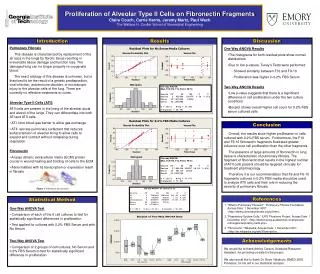
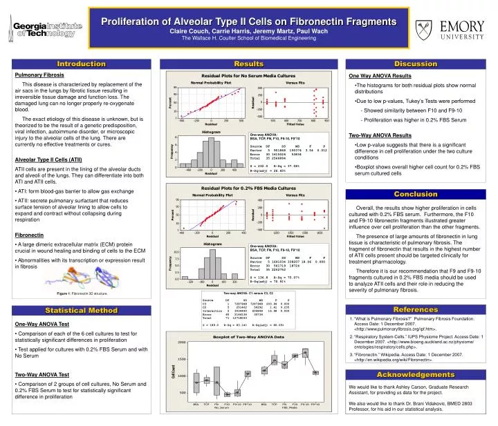
Proliferation of Alveolar Type II Cells on Fibronectin Fragments Claire Couch, Carrie Harris, Jeremy Martz, Paul Wach The Wallace H. Coulter School of Biomedical Engineering Introduction Results Discussion • Pulmonary Fibrosis • This disease is characterized by replacement of the air sacs in the lungs by fibrotic tissue resulting in irreversible tissue damage and function loss. The damaged lung can no longer properly re-oxygenate blood. • The exact etiology of this disease is unknown, but is theorized to be the result of a genetic predisposition, viral infection, autoimmune disorder, or microscopic injury to the alveolar cells of the lung. There are currently no effective treatments or cures. • Alveolar Type II Cells (ATII) • ATII cells are present in the lining of the alveolar ducts and alveoli of the lungs. They can differentiate into both ATI and ATII cells. • ATI: form blood-gas barrier to allow gas exchange • ATII: secrete pulmonary surfactant that reduces surface tension of alveolar lining to allow cells to expand and contract without collapsing during respiration • Fibronectin • A large dimeric extracellular matrix (ECM) protein crucial in wound healing and binding of cells to the ECM • Abnormalities with its transcription or expression result in fibrosis • One Way ANOVA Results • The histograms for both residual plots show normal distributions • Due to low p-values, Tukey’s Tests were performed • - Showed similarity between F10 and F9-10 • - Proliferation was higher in 0.2% FBS Serum • Two-Way ANOVA Results • Low p-value suggests that there is a significant difference in cell proliferation under the two culture conditions • Boxplot shows overall higher cell count for 0.2% FBS serum cultured cells One-way ANOVA: BSA, TCP, FN, F10, F9-10, F9*10 Source DF SS MS F P Factor 5 951868 190374 3.54 0.012 Error 30 1615026 53834 Total 35 2566894 S = 232.0 R-Sq = 37.08% R-Sq(adj) = 26.60% Conclusion Overall, the results show higher proliferation in cells cultured with 0.2% FBS serum. Furthermore, the F10 and F9-10 fibronectin fragments illustrated greater influence over cell proliferation than the other fragments. The presence of large amounts of fibronectin in lung tissue is characteristic of pulmonary fibrosis. The fragment of fibronectin that results in the highest number of ATII cells present should be targeted clinically for treatment pharmacology. Therefore it is our recommendation that F9 and F9-10 fragments cultured in 0.2% FBS media should be used to analyze ATII cells and their role in reducing the severity of pulmonary fibrosis. One-way ANOVA: BSA, TCP, FN, F10, F9-10, F9*10 Source DF SS MS F P Factor 5 1691034 338207 18.06 0.000 Error 30 561719 18724 Total 35 2252752 S = 136.8 R-Sq = 75.07% R-Sq(adj) = 70.91% Figure 1: Fibronectin 3D structure. Two-way ANOVA: C1 versus C3, C2 Source DF SS MS F P C3 1 7287948 7287948 203.94 0.000 C2 5 251462 50292 1.41 0.235 Interaction 5 3034490 606898 16.98 0.000 Error 60 2144134 35736 Total 71 12718033 S = 189.0 R-Sq = 83.14% R-Sq(adj) = 80.05% References Statistical Method • “What is Pulmonary Fibrosis?” Pulmonary Fibrosis Foundation. Access Date: 1 December 2007. <http://www.pulmonaryfibrosis.org/ipf.htm>. • “Respiratory System-Cells.” IUPS Physiome Project. Access Date: 1 December 2007. <http://www.bioeng.auckland.ac.nz/physiome/ ontologies/respiratory/cells.php>. • “Fibronectin.” Wikipedia. Access Date: 1 December 2007. <http://en.wikipedia.org/wiki/Fibronectin>. • One-Way ANOVA Test • Comparison of each of the 6 cell cultures to test for statistically significant differences in proliferation • Test applied for cultures with 0.2% FBS Serum and with No Serum • Two-Way ANOVA Test • Comparison of 2 groups of cell cultures, No Serum and 0.2% FBS Serum to test for statistically significant difference in proliferation Acknowledgements We would like to thank Ashley Carson, Graduate Research Assistant, for providing us data for the project. We also would like to thank Dr. Brani Vidakovic, BMED 2803 Professor, for his aid in our statistical analysis.