Download
1 / 71
900 likes | 1.43k Vues
Chapter 3: Amino Acids, Peptides, and Proteins. Dr. Clower Chem 4202. Outline (part I). Sections 3.1 and 3.2 Amino Acids Chemical structure Acid-base properties Stereochemistry Non-standard amino acids Formation of Peptide Bonds. R side chain | H 2 N— C —COOH | H.

E N D
Chapter 3: Amino Acids, Peptides, and Proteins Dr. Clower Chem 4202
Outline (part I) • Sections 3.1 and 3.2 • Amino Acids • Chemical structure • Acid-base properties • Stereochemistry • Non-standard amino acids • Formation of Peptide Bonds
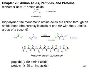
R side chain • | • H2N— C —COOH| • H Amino Acids • The building blocks of proteins • Also used as single molecules in biochemical pathways • 20 standard amino acids (a-amino acids) • Two functional groups: • carboxylic acid group • amino group on the alpha () carbon • Have different side groups (R) • Properties dictate behavior of AAs
Zwitterions • Both the –NH2 and the –COOH groups in an amino acid undergo ionization in water. • At physiological pH (7.4), a zwitterion forms • Both + and – charges • Overall neutral • Amphoteric • Amino group is protonated • Carboxyl group is deprotonated • Soluble in polar solvents due to ionic character • Structure of R also influence solubility
Classification of Amino Acids • Classify by structure of R • Nonpolar • Polar • Aromatic • Acidic • Basic
Nonpolar Amino Acids • Hydrophobic, neutral, aliphatic
Polar Amino Acids • Hydrophilic, neutral, typically H-bond
Disulfide Bonds • Formed from oxidation of cysteine residues
Aromatic Amino Acids • Bulky, neutral, polarity depend on R
Acidic R group = carboxylic acid Donates H+ Negatively charged Basic R group = amine Accepts H+ Positively charged His ionizes at pH 6.0 Acidic and Basic Amino Acids
pK1 ~ 2.2 (protonated below 2.2) pK2 ~ 9.4 (NH3+ below 9.4) pKR (when applicable) Acid-base Properties • Remember H3PO4 (multiple pKa’s) • AAs also have multiple pKa’s due to multiple ionizable groups
Note 3-letter and 1-letter abbreviations Table 3-1 Amino acid organization chart
pH and Ionization • Consider glycine: • Note that the uncharged species never forms
Titration of Glycine • pK1 • [cation] = [zwitterion] • pK2 • [zwitterion] = [anion] • First equivalence point • Zwitterion • Molecule has no net charge • pH = pI (Isoelectric point) • pI = average of pKa’s = ½ (pK1 + pK2) • pIglycine = ½ (2.34 + 9.60) = 5.97 • Animation
pI of Lysine • For AAs with 3 pKa’s, pI = average of two relevant pKa values • Consider lysine (pK1 = 2.18, pK2 = 8.95, pKR = 10.53): • Which species is the isoelectric form? • So, pI = ½ (pK2 + pKR) = ½ (8.95 + 10.53) = 9.74 • Note: pKR is not always higher than pK2 (see Table 3-1 and Fig. 3-12)
Learning Check • Would the following ions of serine exist at a pH above, below, or at pI?
Stereochemistry of AAs • All amino acids (except glycine) are optically active • Fischer projections:
D and L Configurations • d = dextrorotatory • l = levorotatory • D, L = relative to glyceraldehyde
Importance of Stereochemistry • All AA’s found in proteins are L geometry • S enantiomer for all except cysteine • D-AA’s are found in bacteria • Geometry of proteins affects reactivity (e.g binding of substrates in enzymes) • Thalidomide
Non-standard Amino Acids • AA derivatives • Modification of AA after protein synthesized • Terminal residues or R groups • Addition of small alkyl group, hydroxyl, etc. • D-AAs • Bacteria
CHEM 2412 Review • Carboxylic acid + amine = ? • Structure of amino acid
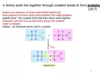
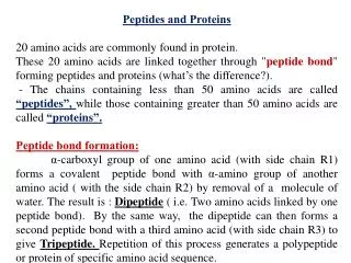
The Peptide Bond • Chain of amino acids = peptide or protein • Amino acid residues connected by peptide bonds • Residue = AA – H2O
Rigid restricted rotation The Peptide Bond • Non-basic and non-acidic in pH 2-12 range due to delocalization of N lone pair • Amide linkage is planar, NH and CO are anti
Polypeptides • Linear polymers (no branches) • AA monomers linked head to tail • Terminal residues: • Free amino group (N-terminus) • Draw on left • Free carboxylate group (C-terminus) • Draw on right • pKa values of AAs in polypeptides differ slightly from pKa values of free AAs
Naming Peptides • Name from the free amine (NH3+) • Use -yl endings for the names of the amino acids • The last amino acid with the free carboxyl group (COO-) uses its amino acid name (GA)
Amino Acid Ambiguity • Glutamate (Glu/E) vs. Glutamine (Gln/Q) • Aspartate (Asp/D) vs. Asparagine (Asn/N) • Converted via hydrolysis • Use generic abbreviations for either • Glx/Z • Asx/B • X = undetermined or nonstandard AA
Learning Check Write the name of the following tetrapeptide using amino acid names and three-letter abbreviations.
Learning Check • Draw the structural formula of each of the following peptides. A. Methionylaspartic acid B. Alanyltryptophan C. Methionylglutaminyllysine D. Histidylglycylglutamylalanine
Outline (part II) • Sections 3.3 and 3.4 • Separation and purification • Protein sequencing • Analysis of primary structure
Protein size • In general, proteins contain > 40 residues • Minimum needed to fold into tertiary structure • Usually 100-1000 residues • Percent of each AA varies • Proteins separated based on differences in size and composition • Proteins must be pure to analyze, determine structure/function
Factors to control • pH • Keep pH stable to avoid denaturation or chemical degradation • Presence of enzymes • May affect structure (e.g. proteases/peptidase) • Temperature • Control denaturation (0-4°C) • Control activity of enzymes • Thiol groups • Reactive • Add protecting group to prevent formation of new disulfide bonds • Exposure to air, water • Denature or oxidize • Store under N2 or Ar • Keep concentration high
General Separation Procedure • Detect/quantitate protein (assay) • Determine a source (tissue) • Extract protein • Suspend cell source in buffer • Homogenize • Break into fine pieces • Cells disrupted • Soluble contents mix with buffer • Centrifuge to separate soluble and insoluble • Separate protein of interest • Based on solubility, size, charge, or binding ability
Solubility • Selectively precipitate protein • Manipulate • Concentration of salts • Solvent • pH • Temperature
Concentration of salts • Adding small amount of salt increases [Protein] • Salt shields proteins from each other, less precipitation from aggregation • Salting-in • Salting out • Continue to increase [salt] decreases [protein] • Different proteins salt out at different [salt]
Other Solubility Methods • Solvent • Similar theory to salting-out • Add organic solvent (acetone, ethanol) to interact with water • Decrease solvating power • pH • Proteins are least soluble at pI • Isoelectric precipitation • Temperature • Solubility is temperature dependent
Chromatography • Mobile phase • Mixture dissolved in liquid or solid • Stationary phase • Porous solid matrix • Components of mixture pass through the column at different rates based on properties
Types of Chromatography • Paper • Stationary phase = filter paper • Same theory as thin layer chromatography (TLC) • Components separate based on polarity • High-performance liquid (HPLC) • Stationary phase = small uniform particles, large surface area • Adapt to separate based on polarity, size, etc. • Hydrophobic Interaction • Hydrophobic groups on matrix • Attract hydrophobic portions of protein
Types of Chromatography • Ion-exchange • Stationary phase = chemically modified to include charged groups • Separate based on net charge of proteins • Anion exchangers • Cation groups (protonated amines) bind anions • Cation exchangers • Anion groups (carboxylates) bind cations
Types of Chromatography • Gel-filtration • Size/molecular exclusion chromatography • Stationary phase = gels with pores of particular size • Molecules separate based on size • Small molecules caught in pores • Large molecules pass through
Types of Chromatography • Affinity • Matrix chemically altered to include a molecule designed to bind a particular protein • Other proteins pass through
UV-Vis Spectroscopy • Absorbance used to monitor protein concentrations of each fraction • l = 280 nm • Absorbance of aromatic side groups
Electrophoresis • Migration of ions in an electric field • Electrophoretic mobility (rate of movement) function of charge, size, voltage, pH • The positively charged proteins move towards the negative electrode (cathode) • The negatively charged proteins move toward the positive electrode (anode) • A protein at its pI (neutral) will not migrate in either direction • Variety of supports (gel, paper, starch, solutions)
Protein Sequencing • Determination of primary structure • Need to know to determine 3D structure • Gives insight into protein function • Approach: • Denature protein • Break protein into small segments • Determine sequences of segments • Animation
End group analysis • Identify number of terminal AAs • Number of chains/subunits • Identify specific AA • Dansyl chloride/dabsyl chloride • Sanger method (FDNB) • Edman degradation (PITC) Bovine insulin
Dansyl chloride • Reacts with primary amines • N-terminus • Yields dansylated polypeptides • Dansylated polypeptides hydrolyzed to liberate the modified dansyl AA • Dansyl AA can be identified by chromatography or spectroscopy (yellow fluorescence) • Useful method when protein fragmented into shorter polypeptides
Dabsyl chloride and FDNB • Same result as dansyl chloride • Dabsyl chloride • 1-Fluoro-2,4-dinitrobenzene (FDNB) • Sanger method
Edman degradation • Phenylisothiocyanate (PITC) • Reacts with N-terminal AA to produce a phenylthiocarbamyl (PTC) • Treat with TFAA (solvent/catalyst) to cleave N-terminal residue • Does not hydrolyze other AAs • Treatment with dilute acid makes more stable organic compound • Identify using NMR, HPLC, etc. • Sequenator (entire process for proteins < 100 residues)
Fragmenting Proteins • Formation of smaller segments to assist with sequencing • Process: • Cleave protein into specific fragments • Chemically or enzymatically • Break disulfide bonds • Purify fragments • Sequence fragments • Determine order of fragments and disulfide bonds
Cleaving Disulfide Bonds • Oxidize with performic acid • Cys residues form cysteic acid • Acid can oxidize other residues, so not ideal
Cleaving Disulfide Bonds • Reduce by mercaptans (-SH) • 2-Mercaptoethanol • HSCH2CH2OH • Dithiothreitol (DTT) • HSCH2CH(OH)CH(OH)CH2SH • Reform cysteine residues • Oxidize thiol groups with iodoacetete (ICH2CO2-) to prevent reformation of disulfide bonds