Download
1 / 29
320 likes | 525 Vues
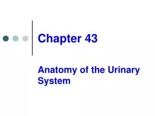
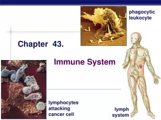
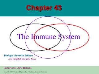
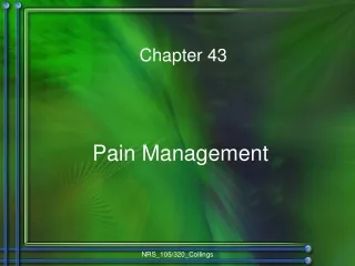
Chapter 43. The Immune System. 3 m. Figure 43.1 A macrophage (blue) ingesting a yeast cell (green). INNATE IMMUNITY Rapid responses to a broad range of microbes. ACQUIRED IMMUNITY Slower responses to specific microbes. External defenses. Internal defenses. Skin. Phagocytic cells.

E N D
Chapter 43 The Immune System
3m Figure 43.1 A macrophage (blue) ingesting a yeast cell (green)
INNATE IMMUNITY Rapid responses to a broad range of microbes ACQUIRED IMMUNITY Slower responses to specific microbes External defenses Internal defenses Skin Phagocytic cells Humoral response (antibodies) Mucous membranes Antimicrobial proteins Secretions Inflammatory response Invading microbes (pathogens) Cell-mediated response (cytotoxic lymphocytes) Natural killer cells Figure 43.2 Overview of vertebrate defenses against bacteria, viruses, and other pathogens
10m Figure 43.3 External innate defense by mucous membranes
Pseudopodia surround microbes. 1 Microbes Microbes are engulfed into cell. 2 MACROPHAGE Vacuole containing microbes forms. 3 Lysosome containing enzymes Vacuole Vacuole and lysosome fuse. 4 Toxic compounds and lysosomal enzymes destroy microbes. 5 Microbial debris is released by exocytosis. 6 Figure 43.4 Phagocytosis
Interstitial fluid bathing the tissues, along with the white blood cells in it, continually enters lymphatic capillaries. 1 Lymphatic capillary Interstitial fluid Fluid inside the lymphatic capillaries, called lymph, flows through lymphatic vessels throughout the body. 2 Adenoid Tonsil Lymphatic vessels return lymph to the blood via two large ducts that drain into veins near the shoulders. 4 Lymph nodes Blood capillary Spleen Lymphatic vessel Tissue cells Peyer’s patches (small intestine) Within lymph nodes, microbes and foreign particles present in the circulating lymph encounter macro- phages, dendritic cells, and lymphocytes, which carry out various defensive actions. 3 Appendix Lymphatic vessels Masses of lymphocytes and macrophages Lymph node Figure 43.5 The human lymphatic system
Blood clot Pin Pathogen Macrophage Blood clotting elements Chemical signals Phagocytic cells Phagocytosis Capillary Red blood cell Fluid, antimicrobial proteins, and clotting elements move from the blood to the site. Clotting begins. 1 Chemical signals released by activated macrophages and mast cells at the injury site cause nearby capillaries to widen and become more permeable. Neutrophils and macrophages phagocytose pathogens and cell debris at the site, and the tissue heals. Chemokines released by various kinds of cells attract more phagocytic cells from the blood to the injury site. 4 2 3 Figure 43.6 Major events in the local inflammatory response
Antigen- binding sites Epitopes (antigenic determinants) Antibody A Antigen Antibody B Antibody C Figure 43.7 Epitopes (antigenic determinants)
Antigen- binding site Antigen- binding site Antigen- binding site Disulfide bridge V V V Variable regions V Light chain V V C C Constant regions C C C C Transmembrane region Plasma membrane b chain a chain Heavy chains Disulfide bridge T cell B cell Cytoplasm of B cell (a) A B cell receptor consists of two identical heavy chains and two identical light chains linked by several disulfide bridges. (b) • A T cell receptor consists of one • chain and one b chain linked by a disulfide bridge. Figure 43.8 Antigen receptors on lymphocytes Cytoplasm of T cell
Infected cell Antigen- presenting cell 1 1 Microbe 1 A fragment of foreign protein (antigen) inside the cell associates with an MHC molecule and is transported to the cell surface. Antigen fragment Antigen fragment Class II MHC molecule Class I MHC molecule 2 2 2 T cell receptor The combination of MHC molecule and antigen is recognized by a T cell, alerting it to the infection. T cell receptor Helper T cell (a) Cytotoxic T cell (b) Figure 43.9 The interaction of T cells with MHC molecules
Bone marrow Thymus Lymphoid stem cell T cell B cell Blood, lymph, and lymphoid tissues (lymph nodes, spleen, and others) Figure 43.10 Overview of lymphocyte development
V4–V39 DNA of undifferentiated B cell V40 J1 J2 J3 J4 J5 V2 V3 Intron V1 C 4 3 2 1 Deletion of DNA between a V segmentand J segment and joining of the segments DNA of differentiated B cell V3 V1 V2 J5 Intron C Transcription of resulting permanently rearranged,functional gene V3 J5 C pre-mRNA Intron RNA processing (removal of intron; addition of capand poly (A) tail) V3 J5 C Poly (A) mRNA Cap Translation Light-chain polypeptide V C B cell receptor Variable region Constant region B cell Figure 43.11 Immunoglobulin gene rearrangement
Antigen molecules bind to the antigen receptors of only one of the three B cells shown. Antigen molecules B cells that differ in antigen specificity Antigen receptor The selected B cell proliferates, forming a clone of identical cells bearing receptors for the selecting antigen. Some proliferating cells develop into long-lived memory cells that can respond rapidly upon subsequent exposure to the same antigen. Some proliferating cells develop into short-lived plasma cells that secrete antibodies specific for the antigen. Antibody molecules Clone of memory cells Clone of plasma cells Figure 43.12 Clonal selection of B cells
Day 1: First exposure to antigen A Secondary response to anti- gen A produces antibodies to A; primary response to anti- gen B produces antibodies to B 1 4 Day 28: Second exposure to antigen A; first exposure to antigen B 3 Primary response to antigen A produces anti- bodies to A 2 104 103 Antibody concentration (arbitrary units) Antibodies to A 102 Antibodies to B 101 100 21 14 28 42 49 56 0 7 35 Time (days) Figure 43.13 The specificity of immunological memory
Cell-mediated immune response Humoral immune response First exposure to antigen Antigens engulfed and displayed by dendritic cells Activate HelperT cell Gives rise to Active and memory helperT cells Figure 43.14 An overview of the acquired immune response (layer 1)
Cell-mediated immune response Humoral immune response First exposure to antigen Antigens displayedby infected cells Antigens engulfed and displayed by dendritic cells Activate Activate Secreted cytokines activate HelperT cell CytotoxicT cell Gives rise to Gives rise to Active and memory helperT cells Memory cytotoxicT cells Active cytotoxicT cells Defend against infected cells, cancer cells, and transplanted tissues Figure 43.14 An overview of the acquired immune response (layer 2)
Cell-mediated immune response Humoral immune response First exposure to antigen Antigens displayedby infected cells Antigens engulfed and displayed by dendritic cells Intact antigens Activate Activate Activate Secreted cytokines activate B cell HelperT cell CytotoxicT cell Gives rise to Gives rise to Gives rise to Active and memory helperT cells Memory cytotoxicT cells Active cytotoxicT cells MemoryB cells Plasmacells Defend against infected cells, cancer cells, and transplanted tissues Secrete antibodies that defend againstpathogens and toxins in extracellular fluid Figure 43.14 An overview of the acquired immune response (layer 3)
After a dendritic cell engulfs and degrades a bacterium, it displays bacterial antigen fragments (peptides) complexed with a class II MHC molecule on the cell surface. A specific helper T cell binds to the displayed complex via its TCR with the aid of CD4. This interaction promotes secretion of cytokines by the dendritic cell. Cytotoxic T cell Dendritic cell Peptide antigen Helper T cell Cell-mediated immunity (attack on infected cells) Class II MHC molecule Bacterium TCR Humoral immunity (secretion of antibodies by plasma cells) CD4 Dendritic cell Cytokines B cell Proliferation of the T cell, stimulated by cytokines from both the dendritic cell and the T cell itself, gives rise to a clone of activated helper T cells (not shown), all with receptors for the same MHC–antigen complex. The cells in this clone secrete other cytokines that help activate B cells and cytotoxic T cells. 1 3 3 1 2 2 Figurze 43.15 The central role of helper T cells in humoral and cell-mediated immune responses
The granzymes initiate apoptosis within the target cells, leading to fragmentation of the nucleus, release of small apoptotic bodies, and eventual cell death. The released cytotoxic T cell can attack other target cells. A specific cytotoxic T cell binds to a class I MHC–antigen complex on a target cell via its TCR with the aid of CD8. This interaction, along with cytokines from helper T cells, leads to the activation of the cytotoxic cell. The activated T cell releases perforin molecules, which form pores in the target cell membrane, and proteolytic enzymes (granzymes), which enter the target cell by endocytosis. Cytotoxic T cell Released cytotoxic T cell Perforin Cancer cell Granzymes Apoptotic target cell TCR CD8 Class I MHC molecule Pore Target cell Peptide antigen Cytotoxic T cell 3 1 2 3 1 2 Figure 43.16 The killing action of cytotoxic T cells
Bacterium Macrophage Peptide antigen Class II MHCmolecule CD4 TCR Helper T cell 1 Figure 43.17 Humoral immune response (layer 1)
Bacterium Macrophage Peptide antigen Class II MHCmolecule B cell CD4 TCR Cytokines Helper T cell Activated helper T cell 2 1 Figure 43.17 Humoral immune response (layer 2)
Bacterium Macrophage Peptide antigen Class II MHCmolecule B cell Secreted antibodymolecules Clone of plasma cells CD4 TCR Endoplasmicreticulum of plasma cell Cytokines Helper T cell Activated helper T cell Clone of memoryB cells 3 1 2 Figure 43.17 Humoral immune response (layer 3)
IgM (pentamer) First Ig class produced after initial exposure to antigen; then its concentration in the blood declines Promotes neutralization and agglutination of antigens; very effective in complement activation (see Figure 43.19) J chain Most abundant Ig class in blood; also present in tissue fluids IgG (monomer) Only Ig class that crosses placenta, thus conferring passive immunity on fetus Promotes opsonization, neutralization, and agglutination of antigens; less effective in complement activation than IgM (see Figure 43.19) IgA (dimer) Present in secretions such as tears, saliva, mucus, and breast milk Provides localized defense of mucous membranes by agglutination and neutralization of antigens (see Figure 43.19) Presence in breast milk confers passive immunity on nursing infant J chain Secretory component IgE (monomer) Triggers release from mast cells and basophils of histamine and other chemicals that cause allergic reactions (see Figure 43.20) IgD (monomer) Present primarily on surface of naive B cells that have not been exposed to antigens Acts as antigen receptor in antigen-stimulated proliferation and differentiation of B cells (clonal selection) Transmembrane region Figure 43.18 The five classes of immunoglobulins
Binding of antibodies to antigens inactivates antigens by Viral neutralization (blocks binding to host) and opsonization (increases phagocytosis) Agglutination of antigen-bearing particles, such as microbes Activation of complement system and pore formation Precipitation of soluble antigens Complement proteins Bacteria Virus MAC Pore Soluble antigens Bacterium Foreign cell Leads to Enhances Cell lysis Phagocytosis Macrophage Figure 43.19 Antibody-mediated mechanisms of antigen disposal
Table 43.1 Blood Groups That Can and Cannot Be Safely Combined in Transfusion
IgE Allergen Histamine 1 3 2 Granule Mast cell Degranulation of the cell, triggered by cross-linking of adjacent IgE molecules, releases histamine and other chemicals, leading to allergy symptoms. 1 IgE antibodies produced in response to initial exposure to an allergen bind to receptors or mast cells. 2 3 On subsequent exposure to the same allergen, IgE molecules attached to a mast cell recog- nize and bind the allergen. Figure 43.20 Mast cells, IgE, and the allergic response
Figure 43.21 X-ray of a hand deformed by rheumatoid arthritis
1µm Figure 43.22 A T cell infected with HIV