Download
1 / 82
820 likes | 1.09k Vues
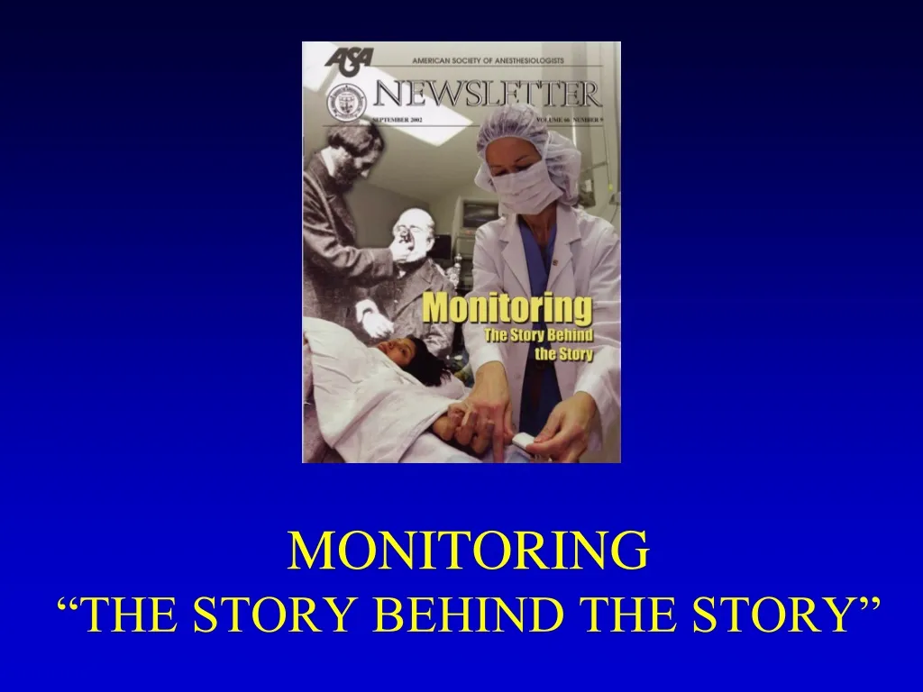
MONITORING “THE STORY BEHIND THE STORY”. Human error…... ASA Status. Arbous MS, Grobbee DE, et al. Anaesthesia 2001;56:1141-53. 869483 anesthesia 769 pts died within 24 hours after anesthesia 42 pts comatose.

E N D
Human error…... ASA Status
Arbous MS, Grobbee DE, et al. Anaesthesia 2001;56:1141-53 869483 anesthesia 769 pts died within 24 hours after anesthesia 42 pts comatose Inadequate monitoring → 10% anesthesia related deaths Postop monitoring: inadequately 8% pts unavailable 5% pts
Arbous MS, Grobbee DE, et al. Anaesthesia 2001;56:1141-53
“Vital Signs” Monitoring Guidelines Cardiovascular Respiratory Others
ASA standards for basic anesthetic monitoring 2001 Standard 1: Qualified anesthesia personnel shall be present in the room throughout the conduct of all general anesthetics, regional anesthetics and monitored anesthesia care Standard 2: During all anesthetics, the patient's oxygenation, ventilation, circulation, and temperature shall be continually evaluated
ASA standards for basic anesthetic monitoring 2001 Oxygenation: Oxygen analyzer for inspired gases-Observation of the patientPulse oximetry Ventilation: Auscultation of breath sounds-Observation of the patientObservation of the reservoir bagCapnography (Carbon dioxide monitoring) Circulation: Continuous ECG displayHeart rate and BP recorded every 5 minutesEvaluation of circulationAuscultation of heart soundsPalpation of pulsePulse plethysmographyPulse oximetryIntrarterial pressure tracing Temperature: Monitor temperature when changes are intended, anticipated, or suspected
Buhre W and Rossaint R. The Lancet 2003; 362:1839-46
Buhre W and Rossaint R. The Lancet 2003; 362:1839-46 Monitoring recommendations of the Association of Anesthetists of Great Britain and Ireland
Buhre W and Rossaint R. The Lancet 2003; 362:1839-46
Buhre W and Rossaint R. The Lancet 2003; 362:1839-46
Buhre W and Rossaint R. The Lancet 2003; 362:1839-46 “There is growing evidence that no single monitoring device can improve outcome in the OR or in the ICU.
ECG blood pressure pulse oximetry capnography + anesthetic gas concentrations + FiO2 temperature
Immediately available Anesthesiology 2002;96:742-52
Periop monitoring Guidelines Cardiovascular Respiratory Others
What to expect from the ECG • Essential monitor • Rate, rhythm, propagation of the excitation wave, heart position, muscle hypertrophy, regional ischemia • NO information about pump function
Lead Selection • Lead II is the same as standard lead two as seen in a 12 lead ECG. • It is the most common monitoring lead. • It is not the optimal monitoring lead.
Lead Selection • V5 = the best lead to detect ST-T change
The shape of the ECG P T QR S
ECG interpretation Rate Rhythm Intervals QRS complexes ST segments & T waves
Intraoperative Monitoring invasive • - invasive AP • Swan Ganz cath • PiCCO System • Advanced PAC: • SVO2, CCO, REF, EDV • …………………TEE
Intraoperative Monitoring invasive
Intraoperative Monitoring invasive 8’ 3’’ + oscill 2-4’’
NIBP (auscultatory / oscillometric) Pros Healthy patients Short case Cons Bladder cuff size Flow dependent Motion Interruption of IV infusion Injury Cuff deflation rate Hydrostatic errors Arterial Cannulation Pros Continuous BP Sick patients Difficult cases ABG monitoring Cons Nerve dysfunction Thrombosis / Ischemia Hematoma formation Infection Hydrostatic errors NIBP vs. Arterial Cannulation
Art Line Complications • Thrombus formation • Arterial laceration • Hematoma • Loss of distal perfusion to hand…ouch! • Nerve dysfunction from dissection • Infection • Errors in monitoring • Failed attempt. Always consider failure as a potential complication.
Arterial Waveform Evaluation • Tf – Foot • Onset of ejection • Systole • T1 - First Shoulder • Peak flow • T2 - Second Shoulder • Peak pressure • Ti – dichotic notch • End of ejection • Closure of aortic valve • Precedes the onset of diastole • Tt – Pulse Duration
Central Line Indications • Peripheral venous access is required for: • Administration of fluids • Administration of drugs • Central venous access is required for: • Parenteral nutrition • Anticipated Inotropic medication infusion • Anticipated large volume resuscitation • Monitoring of central venous pressure (CVP) • Cardiac pacing • Difficult peripheral access
Central Line Contraindications • Patient refusal? • Severe Coagulopathy • Bundle Branch Blocks relative contraindication • Infection at site • Previous failed attempts at specific site • Hematoma • Unusual anatomy
Central Line Techniques • Sterile techniques should be used for all central line cannulation • Surgical scrub with Sterile gown and gloves • Sterile prep of skin and surgical drapes. • Local anesthetic should be used for central catheters in awake patients • Success may be improved by using ultrasound guidance • Techniques of gaining access include: • Catheter over needle • Catheter through needle • Seldinger technique • Surgical cut-down is surgical technique as last resort.
Anatomy of Central Assess • Internal jugular vein • Right sided access preferred. Why? • Apical pleura does not rise as high on right and avoids thoracic duct • Patient positioned head down • In the low approach triangle formed by two heads of sternomastoid and clavicle identified • Cannula aimed down and lateral towards ipsilateral nipple • Subclavian vein • Usually approached from below clavicle • Patient positioned head down • Needle inserted below junction of medial 2/3 and lateral 1/3 of the clavicle • Needle aimed towards suprasternal notch • Passes immediately behind clavicle • Vein encountered after 4-5 cm
Waveform Interpretation • + a wave : This wave is due to the increased atrial pressure during right atrial contraction. It correlates with the P wave on an EKG. • + c wave : This wave is caused by a slight elevation of the tricuspid valve into the right atrium during early ventricular contraction. It correlates with the end of the QRS segment on an EKG. • - x descent : This wave is probably caused by the downward movement of the ventricle during systolic contraction. It occurs before the T wave on an EKG.
Waveform Interpretation • + v wave : This wave arises from the pressure produced when the blood filling the right atrium comes up against a closed tricuspid valve. It occurs as the T wave is ending on an EKG. • - y descent : This wave is produced by the tricuspid valve opening in diastole with blood flowing into the right ventricle. It occurs before the P wave on an EKG.
Periop monitoring Guidelines Cardiovascular Respiratory Others
Monitoring Devices(Types) Stethoscope Used with placement of the endotracheal (ET) tube Will hear breath sounds clearly with the delivery of oxygen into the ET tube with correct placement Can use in placement of nasogastric (NG) tube
Respiratory monitoring AwP (peak-plateau) Peep compliance P/V e flow slope Fi-FeO2 Fi-Fe volatile an. SpO2 EtCO2
AwP (peak-plateau) Peep compliance P/V e flow slope Fi-FeO2 Fi-Fe volatile an. SpO2 EtCO2
During observation in the recovery room, the incidence of hypoxemia in the pulse oximetry group was 1.5-3 time less.
AwP (peak-plateau) Peep compliance P/V e flow slope Fi-FeO2 Fi-Fe volatile an. SpO2 EtCO2
EtCO2CAPNOGRPHY mmHg 80- 40- 0- Time in sec
Tetevossian RG, Wo CC, Shoemaker WC. Crit Care Med 2000;28:2248-53 48 pts whit blunt and hemodynamic instability
Tetevossian RG, Wo CC, Shoemaker WC. Crit Care Med 2000;28:2248-53 PtcO2 and PtCO2 early indicators of tissue hypoxia, subclinical hypovolemia, and hemodynamic shock in ER severely ill patients. Ptc-gas values reflect local skin perfusion during normal conditions and in period of circulatory dysfunction and shock.
Periop monitoring Guidelines Cardiovascular Respiratory Others: Temperature Depth of GA NMT Glycemia Lactate


