Download
1 / 69
700 likes | 1.09k Vues
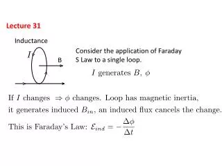
Lecture 31. Quiz on Wed (today): fatty acid synthesis Final on Monday morning 8-10AM. Glycosylphosphatidylinositol (GPI). Anchor proteins to the exterior of the eukaryotic membrane. Alternative to transmembrane polypeptides

E N D
Lecture 31 • Quiz on Wed (today): fatty acid synthesis Final on Monday morning 8-10AM
Glycosylphosphatidylinositol (GPI) • Anchor proteins to the exterior of the eukaryotic membrane. • Alternative to transmembrane polypeptides • Proteins destined to be anchored to the surface of the membrane are synthesized with membrane spanning C-terminal sequences which are removed after GPI addition.
Figure 11–20 Plasma membrane proteins have a variety of functions.
Figure 9.19 Passive transport of solute molecules through a permeable membrane
Figure 9.23 Glucose permease of erythrocyte membrane Passive transport system - intracellular [glucose] =< plasma [glucose] concentration. Also transports epimers of glucose - mannose, galactose at slower rates (20%)
Figure 9.23 Transport of sodium and potassium ions by Na+-K+ ATPase transporter (pump) Three Na+ ions are transported out of the cell for every two K+ that move inside
Lipases • How are lipids accessed for energy production? • Know the differences between the triacylglycerol lipase and phospholipase A2 mechanisms. • Triacylglycerol lipase uses a catalytic triad similar to Ser proteases (Asp, His, Ser) • Phospholipase A2 uses a catalytic triad but substitutes water for Ser.
Figure 25-3a Substrate binding to phospholipase A2. (a) A hypothetical model of phospholipase A2 in complex with a micelle of lysophosphatidylethanolamine. Page 911
Figure 25-4a The X-ray structure of porcine phospholipase A2 (lavender) in complex with the tetrahedral intermediate mimic MJ33. Page 912
Figure 25-4b The catalytic mechanism of phospholipase A2. What other mechanism does this look like? What are the differences? Page 912
Fatty acid binding proteins • Fatty acids form complexes with intestinal fatty acid-binding protein (I-FABP) which makes them more soluble. • Chylomicrons-transport exogenous (dietary) triacylglycerols and chloestorl packaged into lipoprotein molecules from the intestine to the tissues. • Chylomicrons are released into the bloodstream via transport proteins named for their density. • VLDL (very low density lipoproteins), LDL (low density lipoproteins) - transport endogenous (internally produced) triacylglycerols and cholesterols from the liver to tissues “Bad” • HDL (high density lipoproteins), - transport endogenous cholesterol from the tissues to liver - “Good”
Table 12-6 Characteristics of the Major Classes of Lipoproteins in Human Plasma. Page 439
Lipoprotein lipase • Hydrolyzes triacylglycerol components fo chylomicrons and VLDL to free fatty acids and glycerol. • Fatty acids taken up by tissues. • Glycerol is returned to liver or kidneys to convert to DHAP
Fatty acid oxidation • Once fatty acids are taken into the cell they undergo a series of oxidations to yield energy. • In eukaryotes, occurs in the matrix of mitochondria (same place as TCA cycle) • Triglycerides found in fat cells (adipocytes) or in cytoplasm. • Was found by Knoop back in the day (1904) through the following experiment that the oxidation of the carbon atom to the carboxyl-group is involved in fatty acid breakdown.
Figure 25-8 Franz Knoop’s classic experiment indicating that fatty acids are metabolically oxidized at their b-carbon atom. Page 914
Beta Oxidation of Fatty Acids • Process by which fatty acids are degraded by removal of 2-C units • -oxidation occurs in the mitochondria matrix • The 2-C units are released as acetyl-CoA, not free acetate • The process begins with oxidation of the carbon that is "beta" to the carboxyl carbon, so the process is called"beta-oxidation"
Fatty acid oxidation • Triglycerides are broken down into free fatty acids in the cytoplasm. • Beta-oxidation takes place in the mitochondrial matrix. • Fatty acids must be imported into the matrix • Requires activation of fatty acids in the cytosol (fatty acids converted to acyl-CoA form) • Activated fatty acids (acyl-CoA form) are imported into the mitochondrion.
Fatty acids must first be activated by formation of acyl-CoA • Acyl-CoA synthetase condenses fatty acids with CoA, with simultaneous hydrolysis of ATP to AMP and PPi • Formation of a CoA ester is expensive energetically • Reaction just barely breaks even with ATP hydrolysis Go’ATP hydroysis = -32.3 kJ/mol, Go’ Acyl-CoA synthesis +31.5 kJ/mol. • But subsequent hydrolysis of PPi drives the reaction strongly forward (Go’ –33.6 kJ/mol) Fatty acid + CoA + ATP acyl-CoA + AMP + PPi
Import of acyl-CoA into mitochondria • -oxidation occurs in the mitochondria, requires import of long chain acyl-CoAs • Acyl-CoAs are converted to acyl-carnitines by carnitine acyltransferases. • A translocator then imports Acyl carnitine into the matrix while simultaneously exporting free carnitine to the cytosol • Acyl-carnitine is then converted back to acyl-CoA in the matrix
Figure 25-10 Acylation of carnitine catalyzed by carnitine palmitoyltransferase. Page 915
Figure 25-11 Transport of fatty acids into the mitochondrion. Page 916
Import of acyl-CoA into mitochondria • The acyl group of cytosolic acyl-CoA is transferred to carnitine-releases CoA to cytosol. • Acyl-carnitine is transported into the matrix by the carnitine carrier protein • The acyl group is transferred to a CoA molecule from the mitochondrion. • Carnitine is returned to the cytosol.
Deficiencies of carnitine or carnitine transferase or translocator activity are related to disease state • Symptons include muscle cramping during exercise, severe weakness and death. • Affects muscles, kidney, and heart tissues. • Muscle weakness related to importance of fatty acids as long term energy source • People with this disease supplement diet with medium chain fatty acids that do not require carnitine shuttle to enter mitochondria.
Activation of fatty acids for -oxidation • Activation of fatty acid to acyl-CoA form by acyl-CoA synthetase-requires CoASH and ATP (converted to AMP + PPi ) in the cytosol. • Acyl-CoA is converted to acyl-carnitine by carnitine acyltransferase (carnitine palmitoyl transferase I) in the cytosol for transport into the mitochondrion. • Acyl-carnitine is transported across the membrane by the carnitine carrier protein. • Acyl-carnitine is converted to acyl-CoA by carnitine palmitoyl transferase II in the mitochondrial matrix. • The fatty acyl-CoA is ready for the reactions of the oxidation pathway
-2OC-C=C-CO2- -oxidation -2OC-CH2-CH2-CO2- Succinate FAD Succinate dehydrogenase • The first 3 steps resemble the citric acid reactions that convert succinate to oxaloacetate. FADH2 H Fumarate H2O H Fumarase OH Malate -2OC-CH2-C-CO2- NAD+ Malate dehydrogenase NADH + H+ O -2OC-CH2-C-CO2- Oxaloacetate
Formation of a trans double bond by dehydrogenation by acyl-CoA dehydrogenase (AD). • Hydration of the double bond by enoyl-CoA hydratase (EH) to form 3-L-hydroxyacyl-CoA • NAD+-dependent dehydrogenation of b-hydroxyacyl-CoA by 3-L-hydroxyacyl-CoA dehydrogense (HAD) to form -ketoacyl-CoA. • C-C bond cleavage by -ketoacyl-CoA thiolase (KT) Page 917
b-oxidation • Strategy: create a carbonyl group on the-C • First 3 reactions do that; fourth cleaves the "-keto ester" in a reverse Claisen condensation • Products: an acetyl-CoA and a fatty acid two carbons shorter
Acyl-CoA Dehydrogenase • Oxidation of the C-Cbond • Mechanism involves proton abstraction, followed by double bond formation and hydride removal by FAD • Electrons are passed to an electron transfer flavoprotein (ETF), and then to the electron transport chain.
Acyl-CoA dehydrogenase • Mitochondria have four acyl-CoA dehydrogenases • Specificities for short (C4 to C6), medium (C6 to C10), long (C8-C12), very long (C12 to C18) chain fatty acyl-CoAs. • Reoxidized via the Electron Transport Chain.
Figure 25-13 Ribbon diagram of the active site region in a subunit of medium-chain acyl-CoA dehydrogenase from pig liver mitochondria in complex with octanoyl-CoA. FAD = green Octonoyl=blue CoA =white Glu376 -red General base Page 917
Acyl-CoA Dehydrogenase Net: 2 ATP/2 e- transferred
Formation of a trans double bond by dehydrogenation by acyl-CoA dehydrogenase (AD). • Hydration of the double bond by enoyl-CoA hydratase (EH) to form 3-L-hydroxyacyl-CoA • NAD+-dependent dehydrogenation of b-hydroxyacyl-CoA by 3-L-hydroxyacyl-CoA dehydrogense (HAD) to form -ketoacyl-CoA. • C-C bond cleavage by -ketoacyl-CoA thiolase (KT) Page 917
Enoyl-CoA Hydratase • aka crotonases • Adds water across the double bond • Uses substrates with trans-D2-and cisD2double bonds (impt in b-oxidation of unsaturated FAs) • With trans-D2 substrate forms L-isomer, withcisD2 substrate forms D-isomer. • Normal reaction converts trans-enoyl-CoA to L--hydroxyacyl-CoA
Formation of a trans double bond by dehydrogenation by acyl-CoA dehydrogenase (AD). • Hydration of the double bond by enoyl-CoA hydratase (EH) to form 3-L-hydroxyacyl-CoA • NAD+-dependent dehydrogenation of b-hydroxyacyl-CoA by 3-L-hydroxyacyl-CoA dehydrogense (HAD) to form -ketoacyl-CoA. • C-C bond cleavage by -ketoacyl-CoA thiolase (KT) Page 917
Hydroxyacyl-CoA Dehydrogenase • Oxidizes the-Hydroxyl Group to keto group • This enzyme is completely specific for L-hydroxyacyl-CoA • D-hydroxylacyl-isomers are handled differently • Produces one NADH
Formation of a trans double bond by dehydrogenation by acyl-CoA dehydrogenase (AD). • Hydration of the double bond by enoyl-CoA hydratase (EH) to form 3-L-hydroxyacyl-CoA • NAD+-dependent dehydrogenation of -hydroxyacyl-CoA by 3-L-hydroxyacyl-CoA dehydrogense (HAD) to form -ketoacyl-CoA. • C-C bond cleavage by -ketoacyl-CoA thiolase (KT) Page 917
Thiolase • Nucleophillic sulfhydryl group of CoA-SH attacks the -carbonyl carbon of the 3-keto-acyl-CoA. • Results in the cleavage of the C-Cbond. • Acetyl-CoA and an acyl-CoA (-) 2 carbons are formed
Figure 25-15 Mechanism of action of b-ketoacyl-CoA thiolase. • An active site thiol is added to the substrate b-keto group. • C-C bond cleavage forms an acetyl-CoA carbanion intermediate (Claisen ester cleavage) • The acetyl-CoA intermediate is protonated by an enzyme acid group (acetyl-CoA released) • CoA binds to the enzyme-thioester intermediate • Acyl-CoA is released. Net reaction reduces fatty acid by 2C and acyl-CoA group is free to pass through the cyle again. Page 919
Formation of a trans double bond by dehydrogenation by acyl-CoA dehydrogenase (AD). • Hydration of the double bond by enoyl-CoA hydratase (EH) to form 3-L-hydroxyacyl-CoA • NAD+-dependent dehydrogenation of -hydroxyacyl-CoA by 3-L-hydroxyacyl-CoA dehydrogense (HAD) to form -ketoacyl-CoA. • C-C bond cleavage by -ketoacyl-CoA thiolase (KT) Page 917
b-oxidation • Each round of -oxidation produces 1 NADH, 1 FADH2 and 1 acetyl-CoA. • -oxidation of palmitate (C16:0) yields 129 molecules of ATP • C 16:0-CoA + 7 FAD + 7 NAD+ + 7 H2O + 7 CoA 8 acetyl-CoA + 7 FADH2 + 7 NADH + 7 H+ • Acetyl-CoA = 8 GTP, 24 NADH, 8 FADH2 • Total = 31 NADH = 93 ATPs + 15 FADH2 = 30 ATPs • 2 ATP equivalents (ATP AMP + PPi, PPi 2 Pi) consumed during activation of palmitate to acyl-CoA • Net yield = 129 ATPs
Beta-oxidation of unsaturated fatty acids • Nearly all fatty acids of biological origin have cisdouble bonds between C9 and C10 (9 or 9-double bond). • Additional double bonds occur at 3-carbon intervals (never conjugated). • Examples: oleic acid and linoleic acid. • In linoleic acid one of the double bonds is at an even-numbered carbon and the other double bond is at an odd-numbered carbon atom. • 4 additional enzymes are necessary to deal with these problems. • Need to make cis into trans double bonds
Figure 25-17 Problems in the oxidation of unsaturated fatty acids and their solutions. Page 920
-oxidation of unsaturated fatty acids • -oxidation occurs normally for 3 rounds until a cis-3-enoyl-CoA is formed. • Acyl-CoA dehydrogenase can not add double bond between the and carbons. • Enoyl-CoA isomerase converts this to trans- 2 enoyl-CoA • Now the -oxidation can continue on w/ the hydration of the trans-2-enoyl-CoA • Odd numbered double bonds handled by isomerase
-oxidation of odd chain fatty acids • Odd chain fatty acids are less common • Formed by some bacteria in the stomachs of ruminants and the human colon. • -oxidation occurs pretty much as w/ even chain fatty acids until the final thiolase cleavage which results in a3 carbon acyl-CoA (propionyl-CoA) • Special set of 3 enzymes are required to further oxidize propionyl-CoA • Final Product succinyl-CoA enters TCA cycle
Propionyl-CoA Carboxylase • The first reaction • Tetrameric enzyme that has a biotin prosthetic group • Reactions occur at 2 sites in the enzyme. • Carboxylation of biotin at the N1’ by bicarbonate ion (same as pyruvate carboxylase). Driven by hydrolysis of ATP to ADP and Pi-activates carboxyl group for transfer • Stereospecific transfer of the activated carboxyl group from carboxybiotin to propionyl-CoA to form (S)-methylmalonyl-CoA. Occurs via nucleophillic attack on the carboxybiotin by a carbanion at C2 of propionyl-CoA