Download
1 / 31
350 likes | 459 Vues
Shilpa Sood , MD Richard Rosencrantz, MD New York Medical College, Maria Fareri Children’s Hospital Reviewed by Sunny Hussain , MD of the Professional Education Committee. Viral Hepatitis. Introduction. Hepatitis results from various etiologies:

E N D
ShilpaSood, MD Richard Rosencrantz, MD New York Medical College, Maria Fareri Children’s Hospital Reviewed by Sunny Hussain, MD of the Professional Education Committee Viral Hepatitis
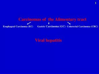
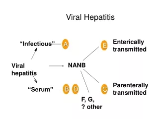
Introduction • Hepatitis results from various etiologies: • Infectious – viral, bacterial, fungal, and parasitic agents. • Non-infectious – drugs, autoimmune, metabolic diseases and ischemic injury. • In the United States, viral hepatitis is most commonly caused by Hepatitis A, Hepatitis B, and Hepatitis C viruses. • Other hepatotropic viruses that cause hepatitis include Hepatitis D and Hepatitis E. • Other and less common causes of viral hepatitis include adenovirus, cytomegalovirus (CMV), Epstein-Barr virus (EBV), and in neonates: herpes simplex virus (HSV).
Case Presentation • 8 year-old male with an ALT of 371 IU/L discovered on routine laboratory testing by the pediatrician. • Denied any abdominal pain, diarrhea, vomiting, rashes, joint pains, fever, jaundice, bruising, and fatigue. No history of chronic medication intake, blood transfusions or known sick contacts. Patient recently immigrated from Haiti few months prior to presentation. • No past medical or surgical history, drug allergies or hospitalizations. • Birth history: born full-term via spontaneous vaginal delivery without perinatal complications, no known prenatal maternal viral screening or perinatal immunizations given. • No family history of gastrointestinal or hepatobiliary disease.
Case Presentation • Physical Exam: well appearing male, vital signs stable, height and weight in the 25-50th %iles. • Examination unremarkable with no stigmata of chronic liver disease. • Labs on the day of evaluation: CBC, complete metabolic panel normal; AST 856 IU/L; ALT 841 IU/L; ALP 420 IU/L; GGT 336 IU/L; total bilirubin 1.4 mg/dL; direct bilirubin: 0.7 mg/dL; total protein 7.9 g/dL; albumin 4.4 g/dL; PT: 11.2s, INR: 1.1, PTT:36s. • Workup: • HAV IgM negative; IgG positive; HBsAg reactive; HBsAb negative; HB core IgM antibody negative; HB core IgG positive; HBeAg reactive; HBeAb negative; HBV DNA >250 million copies/ml; HBV genotype A; HCV antibody negative; HDV Ag negative; EBV VCA IgM 0.27; VCA IgG:1.32; T4/TSH normal; ANA, smooth muscle antibody, LKM antibody negative; no significant serum IgG elevation; HIV non-reactive.
Case Presentation • Liver sonogram: normal size and echotexture of liver with patent vasculature. • Liver biopsy revealed chronic hepatitis consistent with HBV etiology, grade 1 stage 2. • Six months later, surveillance laboratory testing revealed HB-eAg loss with HB-eAb reactivity; however, HBV DNA viral load rose to > 7 billion copies/mL, and a HBV core promoter mutation was detected. • Nucleoside analog therapy was started based on persistently elevated ALT levels 10-20 times the upper limit of normal, log rise in HBV DNA viral load and stage 2 hepatic fibrosis: • 6 months on therapy: transaminases normalized, HBV DNA viral load became undetectable, HB-sAg remained reactive and HBsAb negative. • 1.5 years on therapy: patient remains without signs or symptoms of any chronic liver disease and HCC surveillance has been negative.
Hepatitis A Virus - HAV • Hepatitis A (HAV): • Non-enveloped RNA virus. • Family: • Picornaviridae. • Mode of Transmission: • Person-to-person through fecal-oral contamination. • Incubation period: • 4 weeks. • Clinical features: • Asymptomatic and anicteric to several weeks of malaise, anorexia, nausea, vomiting, and elevated transaminases with or without jaundice. • Prognosis: • No chronic state;10-15% may have a prolonged course. • Isolated HAV infection has excellent prognosis of almost 100% recovery (PALF data).
Management • Treatment: • Supportive management • Prevention: • Hand hygiene. • Post-exposure prophylaxis: • Active immunization with an inactivated Hepatitis A vaccine is recommended for all children in the United States beginning at one year of age; consists of two doses six months apart. • Other indications: unimmunized children travelling to endemic areas (one month before travel), chronic liver disease, receiving clotting factor concentrates, homosexual males and IV drug users. • Passive immunization: HAV-Immunoglobulin is recommended for contacts at day care centers or unimmunized household contacts within two weeks as a single dose. http://www.cdc.gov/hepatitis
Hepatitis B Virus - HBV • Hepatitis B (HBV): • Enveloped DNA virus. • Family: • Hepadnaviridae. • Mode of Transmission: • Parenteral, sexual, and perinatal exposure. • 8 HBV genotypes (A through H), differ in geographic distribution and outcome. • Genotypes A and D: Most common in North America; • B and C are most common in Asia. • Genotype A has most favorable response to treatment compared to other genotypes. • Genotype B when compared with genotype C is more commonly associated with the development of childhood hepatocellular carcinoma (HCC). Yen-Hsuan N et al Gastro 2004
Clinical Features of HBV • Acute HBV infection: • Incubation period: 40-150 days (average 12 weeks). • Prodrome of fever, anorexia, fatigue, malaise and nausea. • 1-2 weeks later, develop jaundice, hepatosplenomegaly, pruritus and intense fatigue that lasts for 1-2 months. • Acute liver failure is rare, occurring in 0.1 - 0.5% of cases. • Chronic HBV: ranges from asymptomatic to symptomatic hepatitis in clinically related phases (see diagram) with progression to cirrhosis with end-stage liver disease in 15-30% and HCC. • Perinatally - acquired infection: • Maternal HB eAg positive – 90% risk. • Maternal HB eAg negative – 20% risk. • Immune tolerant phase is characterized by no signs of significant clinical liver disease. • HB eAg clearance (seroconversion) occurs in approximately 15% by 21 years of age.* http://www.aasld.org/practiceguidelines/Chronic_Hep_B_Update_2009 *Lok ASF et al., Gastro 1987
Clinical Features of HBV • Risk of developing chronic infection decreases with age. • 25% to 30% of infants and children under 5 years of age. • Less than 5% of older children and adults. • Extra hepatic manifestations: • Migratory arthritis, angioedema or maculopapular/urticarial rash, glomerulonephritis, papular acrodermatitis of childhood (Gianotti-Crosti syndrome). • Confirmation of HBV infection is based on serological assays of the specific viral antigens and antibodies. http://www.aasld.org/practiceguidelines/Chronic_Hep_B_Update_2009
Natural History of HBV Infection Elgouhari HM et al. CCJM 2008
Chronic HBV Infection: Phases • Immune tolerance phase: • HB eAg positive, HB eAb negative. • HBV DNA titers high. • Normal LFTs. • Liver biopsy showing minimal or non-specific hepatitis. • Immune clearance phase: • HB eAg positive, HB eAb negative. • HBV DNA titers high. • Elevated LFTs. • Liver biopsy showing chronic hepatitis with moderate or severe chronic necro-inflammation. • Inactive carrier phase: • HB eAg negative, HB eAb positive. • HBV DNA titers low (< 2,000 IU/mL). • Normal LFTs. • Liver biopsy absence of significant hepatitis.
Chronic HBV Infection: Phases • Reactivation phase: • Often associated with precore or core mutations • HB eAg negative, HB eAb positive. • HBV DNA titers moderately high. • Elevated LFTs. • Liver biopsy showing chronic hepatitis. • Resolved hepatitis B: • Previous known history of acute or chronic hepatitis B or presence of HB core IgM negative, IgG positive and HBsAb negative. • HBsAg negative. • Undetectable HBV DNA titers. • Normal ALT levels.
HBV Treatment Algorithm Jonas M, et al, Hepatology 2010
Treatment • Acute HBV: • Supportive care. • Chronic HBV: • Consider therapy if persistently elevated transaminases and liver biopsy-proven significant fibrosis. • Therapeutic options: pegylated-interferon alpha, lamivudine, adefovir dipivoxil, tenofovir, entecavir. • Post-liver transplant: • Long-term HBIG and a nucleoside analog (lamivudine, entecavir, or tenofovir). • YMDD mutation is significant for the development of nucleoside analog resistance. • Immunoprophylaxis: • Prevention of perinatal transmission by active and passive immunization is highly effective. • Infants born to HB sAg positive mothers should receive HBV vaccine and HBIG within 12hrs of birth. http://www.cdc.gov/hepatitis/HBV/VaccChildren.htm
Immunoprophylaxis • HBV vaccine: • Three dose administration for all infants and children under 11 years of age, unimmunized adolescents and adults, high-risk groups such as multiple sexual partners, homosexual males. • Routine post-vaccination HBV serologic testing after the third dose of vaccine (between 9 and 18 months of age at a well baby visit) is advised for infants born to HBs Ag-positive women. • HBV surface antibody testing shouldnotbe performed before nine months of age to avoid confusion with HBIG administered during infancy. http://www.cdc.gov
Hepatitis C virus- HCV • HCV: • Enveloped single-stranded RNA virus. • Family: • Flaviviridae. • Six genotypes (1-6) with subtypes identified differing in treatment response and prognosis. • Genotype 1: • Most common in North America and is associated with less favorable response to treatment (30-50%). • HCV: • Highly prevalent and the most important cause of chronic viral hepatitis in the US. • Most common cause of liver transplantation in adults in US.
Clinical Features • Modes of transmission: • a) Percutaneous: IV drug use, blood trans-fusions, accidental needle stick injury, skin tattooing, • b) Non-percutaneous: intra-familial and sexual routes, risk of vertical transmission is approximately 3-5% (2-3 times higher in HIV/HCV co-infection). • Incubation Period: • 15-50 days with symptoms developing 5-12 weeks after exposure. • Acute HCV infection: • Mostly asymptomatic. • Acute liver failure is rare; however, co-infection with other hepatotropic viruses such as HBV may cause rapid progression of disease.
Clinical Features • Chronic HCV infection: • Spectrum of minimal clinical symptoms to fatigue, jaundice, elevated transaminases. • Extra hepatic manifestations: • Essential mixed cryoglobulinemia, focal lymphocytic sialadenitis, autoimmune thyroiditis, porphyria cutanea tarda, lichen planus and non-Hodgkin lymphoma. • Natural history: • Acute hepatitis C infection chronic hepatitis (85%) cirrhosis (20% over 20 years) HCC (4%) death due to decompensated liver disease (3.6%). • Neonatally acquired infection is associated with a 25% spontaneous clearance rate by 7.3 years.* *Yeung LT et al., J Vital Hepat 2007 Di Bisceglie et al. Hepatology2003
Diagnosis of HCV • Initial test: • Serum HCV antibody • HCV RNA by RT-PCR. • Infants born to HCV infected mothers, • HCV antibody testing performed at 18 months of age or older. • If earlier diagnosis is desired, HCV RNA testing may be considered at 1-2 months of age. • HCV genotyping should be performed prior to treatment to determine prognosis and the duration of therapy.
HCV Clinical Management • Liver biopsy: • Consider for staging and treatment assessment. • Initiating therapy: • Based on the presence of signs and/or symptoms of clinical liver disease, liver biopsy showing severity of liver damage and in some cases parental or patient’s request for therapy. • Cirrhotic patients: • screened for HCC by liver ultrasonography and AFP every 6 months. • Serial Hepatic MR imaging may be used to identify early lesions. • Recurrent HCV infection in patients transplanted for HCV cirrhosis is almost universal.
Treatment Algorithm for HCV Genotype 1 • Acute HCV: • PEG-IFN Monotherapy: • May prevent chronic infection if initiated within 3 months of exposure. • Chronic HCV: • Combination therapy: PEG-IFN + ribavirin. Ghany MG et al, Hepatology 2009
Virologic Response during Therapy • Rapid Virologic Response (RVR): • HCV RNA negative by week 4 of treatment by PCR. • Allows shortening of course for genotypes 2 & 3, and low viral load genotype 1. • Early Virologic Response (EVR): • >2 log reduction in HCV RNA compared to baseline[partial EVR] or HCV RNA negative at week 12 of treatment. • Predicts lack of Sustained Virologic Response (SVR). • Sustained Virologic Response (SVR): • HCV RNA negative 24 weeks after cessation of treatment. • Best predictor of long term response to treatment. Ghany MG et al, Hepatology 2009
Hepatitis D Virus- HDV • Small defective RNA virus, incubation period 35 days. • Dependent on active HBV infection and is associated with more severe clinical liver disease. • Co-infection (simultaneous) or super-infection (pre-existing HBV infection). • Diagnosis: • Acute: HDV antigen + ; either • Co-infection HBV core IgM + • Super-infection: HBV core IgM - • Chronic: HDV antigen - ; HBV core IgM -; HDV RNA+ • More than double the rate of progression to cirrhosis than HBV alone. • Treatment: • No treatment recommendations for chronic HDV in children, rarely interferon-alpha may used.
Hepatitis E Virus- HEV • Family: • Calicivirus. • Mode of transmission: • Fecal-oral route. • Incubation period : • 2-9 weeks. • Affects mainly adolescents and adults. • Clinical Course: • Acute self-limited infection with clinical features similar to HAV; • May be severe in those with preexisting chronic liver disease. • High mortality for pregnant women particularly in the third trimester. • No chronic state.
References • http://www.cdc.gov/hepatitis • Yen-Hsuan N et al Gastro 2004 • http://www.aasld.org/practiceguidelines/Chronic_Hep_B_Update_2009 • Lok ASF et al., Gastro 1987 • http://www.aasld.org/practiceguidelines/Chronic_Hep_B_Update_2009 • Elgouhari HM et al. CCJM 2008 • Jonas M, et al, Hepatology 2010 • Yeung LT et al., J Vital Hepat 2007 • Di Bisceglie et al. Hepatology 2003 • Ghany et al, Hepatology 2009 Achalasia