Download
1 / 38
400 likes | 744 Vues
Acute renal failure. Dr. H. N. Sarker MBBS, FCPS, MACP(USA) Associate professor medicine. Introduction.

E N D
Acute renal failure Dr. H. N. Sarker MBBS, FCPS, MACP(USA) Associate professor medicine
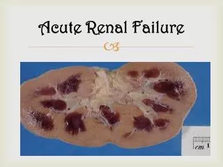
Introduction • Acute renal failure (ARF) refers to a sudden and usually reversible loss of renal function, which develops over a period of days or weeks and is usually accompanied by a reduction in urine volume.
Introduction • Acute renal failure (ARF) is a rapid loss of renal function due to damage to the kidneys, resulting in retention of nitrogenous (urea and creatinine) and non-nitrogenous waste products that are normally excreted by the kidney.
Introduction • Depending on the severity and duration of the renal dysfunction, this accumulation is accompanied by metabolic disturbances, such as metabolic acidosis (acidification of the blood) and hyperkalaemia (elevated potassium levels), changes in body fluid balance, and effects on many other organ systems.
Introduction • It can be characterised by oliguria or anuria (decrease or cessation of urine production), although non-oliguric ARF may occur. It is a life-threatening medical emergency
Causes • Acute renal failure is usually categorised (as in the flowchart below) according to pre-renal, intrinsic and post-renal causes. • Pre-renal Intrinsic Post-renal
Pre-renal (causes in the blood supply): • hypovolemia (decreased blood volume), usually from shock or dehydration and fluid loss or excessive diuretics use. • Heart failure. • hepatorenal syndrome in which renal perfusion is compromised in liver failure • vascular problems, such as atheroembolic disease and renal vein thrombosis (which can occur as a complication of the nephrotic syndrome)
Pre-renal • infection usually sepsis, systemic inflammation due to infection • severe burns • sequestration due to pericarditis and pancreatitis • hypotension due to antihypertensives and vasodilators
Intrinsic (damage to the kidney itself) • Toxins or medication (e.g. some NSAIDs, aminoglycoside antibiotics) • Sepsis • Rhabdomyolysis (breakdown of muscle tissue) • Haemolysis (breakdown of red blood cells)
Post-renal (obstructive causes in the urinary tract ) • benign prostatic hypertrophy or prostate cancer. • kidney stones. • due to abdominal malignancy (e.g. ovarian cancer, colorectal cancer
REVERSIBLE PRE-RENAL ACUTE RENAL FAILURE • Haemodynamic disturbances can initially produce acute renal dysfunction that has the potential to be rapidly reversed, prompt recognition and treatment are important. • These topics are considered separately from established acute renal failure.
Pathogenesis • The kidney can regulate its own blood flow and GFR over a wide range of perfusion pressures. • When the perfusion pressure falls-as in hypovolaemia, shock, heart failure or narrowing of the renal arteries-the resistance vessels in the kidney dilate to facilitate flow. • Vasodilator prostaglandins are important, and this mechanism is markedly impaired by NSAIDs .
Pathogenesis • If autoregulation of blood flow fails, the GFR can still be maintained by selective constriction of the post-glomerular (efferent) arteriole. • This is mediated through the release of renin and generation of angiotensin II, which preferentially constricts this vessel. • ACE inhibitors interfere with this response .
Pathogenesis • More severe or prolonged under-perfusion of the kidneys may lead to failure of these compensatory mechanisms and hence an acute decline in GFR. • The renal tubules are intact and become hyperfunctional; that is, tubular reabsorption of sodium and water is increased.
Pathogenesis • This leads to the formation of a low volume of urine which is concentrated (osmolality > 600 mOsm/kg) but low in sodium (< 20 mmol/l).
Clinical assessment • The clinical picture is often dominated by the underlying condition (e.g. septic shock, trauma,diarrhoea) • There may be marked hypotension and signs of poor peripheral perfusion, such as delayed capillary return.
Clinical assessment • Postural hypotension (a fall in blood pressure > 20/10 mmHg from lying to standing) is a valuable sign of early hypovolaemia. • Metabolic acidosis and hyperkalaemia are often present.
Management • Establish and correct the underlying cause of the ARF. • If hypovolaemia is present, restore blood volume as rapidly as possible (with blood, plasma or isotonic saline (0.9%), depending on what has been lost).
Management • Optimise systemic haemodynamics. Monitoring of the central venous pressure or pulmonary wedge pressure as an adjunct to clinical examination may aid in determining the rate of administration of fluid.
Management • Critically ill patients may require invasive haemodynamic monitoring to assess cardiac output and systemic vascular resistance, and the use of inotropic drugs to restore an effective blood pressure .
Management • Correct metabolic acidosis. • Restoration of blood volume will correct acidosis by restoring kidney function. • Isotonic sodium bicarbonate (e.g. 500 ml of 1.26%) may be used.
ESTABLISHED ACUTE RENAL FAILURE • Established ARF may develop following severe or prolonged under-perfusion of the kidney (pre-renal ARF). In such cases, the histological pattern of acute tubular necrosis is usually seen. • 'renal' and 'post-renal' causes produce established ARF
Features of established ARF • These reflect the causal condition, such as trauma, septicaemia or systemic disease, together with features of renal failure. • 'Uraemic' features -anorexia, nausea and vomiting followed by drowsiness, apathy, confusion, muscle-twitching, hiccoughs, fits and coma.
Features of established of ARF • Alterations in urine volume Patients are usually oliguric (urine volume < 500 ml daily). Anuria (complete absence of urine) is rare and usually indicates acute urinary tract obstruction or vascular occlusion
Features established of ARF • In about 20% of cases, the urine volume is normal or increased (non-oliguric ARF). • Disturbances of water, electrolyte and acid-base balance Hyperkalaemia is common Metabolic acidosis .
Features established of ARF • Hypocalcaemia, due to reduced renal production of 1,25-dihydroxycholecalciferol, is common. • Respiratory rate -increased due to acidosis, pulmonary oedema or respiratory infection. • Anaemia is common, due to excessive blood loss, haemolysis or decreased erythropoiesis .
Diagnosis- Consensus criteria for the diagnosis of ARF are: • Risk: serum creatinine increased 1.5 times OR urine production of <0.5 ml/kg/h body weight for 6 hours • Injury: creatinine 2.0 times OR urine production <0.5 ml/kg/h for 12 h
Diagnosis- • Failure: creatinine 3.0 times OR creatinine >355 μmol/l (with a rise of >44) OR urine output below 0.3 ml/kg/h for 24 h • Loss: persistent ARF or complete loss of kidney function for more than four weeks • End-stage Renal Disease: complete loss of kidney function for more than three months
Diagnosis- Routine investigation- urine for R/E Blood urea & electrolytes S. creatinine ABG
Diagnosis- • Imaging- . Renal ultrasound: usually required urgently to confirm/refute two equal-sized, unobstructed kidneys Chest X-ray, X-ray KUB • ECG: if > 40 years or there are risk factors for cardiac disease
Diagnosis- • Microbiology • Blood cultures • C-reactive protein (ESR is misleading in ARF) • Mid-stream urine • Other cultures, e.g. wound, sputum, catheters • Hepatitis and HIV serology: urgent if dialysis is needed (isolation of dialysis machine if positive)
Management • Emergency resuscitation – • Hyperkalaemia (a plasma K+ concentration > 6 mmol/l) must be treated immediately to prevent the development of life-threatening cardiac arrhythmias. • Circulating blood volume should be optimised to ensure adequate renal perfusion.
Management • Hypovolaemia must be treated as for reversible pre-renal ARF monitoring of central venous or pulmonary wedge pressure as required. • Patients with pulmonary oedema usually require dialysis to remove sodium and water. • Severe acidosis can be ameliorated with isotonic sodium bicarbonate (e.g. 500 ml of 1.26%) if volume status allows.
Management • In an anuric or volume-overloaded patient, renal replacement therapy may be required. • Treatment of the underlying cause of the ARF . • Fluid and electrolyte balance • After initial resuscitation, daily fluid intake should equal urine output, plus an additional 500 ml to cover insensible losses
Management • Protein and energy intake In patients in whom dialysis is likely to be avoided, accumulation of urea is slowed by dietary protein restriction (to about 40 g/day) • Infection control. • Renal replacement therapy
Prognosis • In uncomplicated ARF, such as that due to simple haemorrhage or drugs, mortality is low even when renal replacement therapy is required. • In ARF associated with serious infection and multiple organ failure, mortality is 50-70%.
Prognosis • Outcome is usually determined by the severity of the underlying disorder and other complications, rather than by renal failure itself.