Download
1 / 57
670 likes | 1.35k Vues
Inflammation and Repair. Dr. Maha Arafah. Learning Objectives:. Define inflammation, its causes and clinical appearance. Describe the sequence of vascular changes in acute inflammation ( vasodilation , increased permeability) and their purpose.

E N D
Inflammation and Repair Dr. MahaArafah
Learning Objectives: • Define inflammation, its causes and clinical appearance. • Describe the sequence of vascular changes in acute inflammation (vasodilation, increased permeability) and their purpose. • Know the mechanisms of increased vascular permeability. • Define the terms edema, transudate, and exudate.
Learning Objectives: • Define inflammation, its causes and clinical appearance. • Describe the sequence of vascular changes in acute inflammation (vasodilation, increased permeability) and their purpose. • Know the mechanisms of increased vascular permeability. • Define the terms edema, transudate, and exudate.
What is Inflammation? • It is the way the vascularised living tissue reacts to cell injury. • Aim: eliminate the initial cause of cell injury as well as the necrotic cells and tissues resulting from the original insult. • Cause: Infection, trauma, physical injury, chemical injury, immunologic injury, tissue death.
How does inflammation accomplish the protection? • Inflammation serves to: • destroy, dilute or isolate the injurious agent (microbes, toxins), and • eliminate the necrotic cells and tissues. • Then it starts a series of events which leads as far as possible to the healing and reconstitution of the damaged tissue. Therefore, Inflammation is part of a broader protective response (innate immunity )
Inflammation and repair • During repair, the injured tissue is replaced by: • Regeneration of native parenchyma cells • Filling of the defect by fibroblastic tissue or both • Inflammation and repair are protective response
Can inflammation be harmful ! ? • It can induce harm: e.g. anaphylactic reaction rheumatoid arthritis atherosclerosis
How is inflammation harmful? • The components of the inflammatory reaction that destroy and eliminate microbes and dead tissues are capable of also injuring normal tissues. • This may accompany inflammatory reactions that are: • entirely normal, beneficial (e.g., when the infection is severe), • prolonged (e.g., when the eliciting agent resists eradication) • inappropriate (e.g., when it is directed against self-antigens in autoimmune diseases) • against usually harmless environmental antigens (allergic disorders)
What happens then? • Inflammation is terminated when the offending agent is eliminated and the secreted mediators are broken down or dissipated. • There are active anti-inflammatory mechanisms that serve to control the response and prevent it from causing excessive damage to the host.
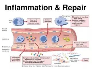
Cells & molecules that play important roles in inflammation • blood leukocytes • plasma proteins • cells of vascular walls • cells of the surrounding connective tissue • extracellular matrix (ECM) of the surrounding connective tissue
Cells & molecules that play important roles in inflammation Cells of surrounding C.T. • The circulating cells: Plasma proteins Blood leukocytes Extracellularmatrix of surrounding C.T. Cells of vascular wall The extracellular matrix
Inflammation is mediated by chemical substances called Chemical Mediators
What is the source of these chemical mediators? • Phagocytes and other host cells • Leukocyte • Endothelium • Mast cell • Plasma proteins
Types of Inflmmation Acute Inflammation Chronic Inflammation is of longer duration associated histologically with the presence of lymphocytes and macrophages, the proliferation of blood vessels, fibrosis, and tissue necrosis. Less uniform. • rapid in onset (seconds or minutes) • relatively short duration, lasting for minutes, several hours, or a few days • its main characteristics: • the exudation of fluid and plasma proteins (edema) • the emigration of leukocytes, predominantly neutrophils.
What are the cardinal signs of inflammation? • Local clinical signs of acute inflammation : • Heat • Redness • Swelling • Pain • Loss of function
What are the steps of the inflammatory response? 5 R’s (1) Recognition of the injurious agent (2) Recruitment of leukocytes (3) Removal of the agent (4) Regulation (control) of the response (5) Resolution
Learning Objectives: • Define inflammation, its causes and clinical appearance 2. Describe the sequence of vascular changes in acute inflammation (vasodilation, increased permeability) and their purpose. 3. Know the mechanisms of increased vascular permeability. 4. Define the terms edema, transudate, and exudate.
Events of acute Inflammation • Acute inflammation is a rapid response to an injurious agent that serves to deliver mediators of host defense-leukocytes and plasma proteins-to the site of injury. • Acute inflammation has three main events: (1) Hemodynamic changes (alterations in vascular caliber that lead to an increase in blood flow) (2) Increased vascular permeability (structural changes in the microvasculature that permit plasma proteins and leukocytes to leave the circulation) (3) Emigration of the leukocytes from the microcirculation (their accumulation in the focus of injury, and their activation to eliminate the offending agent) vascular cellular
(1) Hemodynamic changes (alterations in vascular caliber that lead to an increase in blood flow) (2) Increased vascular permeability Vascular Changes
What are the different phases of changes in Vascular Caliber and Flow?
Phases of changes in Vascular Caliber and Flow • 1. Transient vasoconstriction of arterioles - It disappears within 3-5 seconds in mild injuries - It may last several minutes in more severe injury (burn)
Phases of changes in Vascular Caliber and Flow • 2.Vasodilatation: It involves the arterioles and then results in opening of new microvasculature beds in the area thus leading to increasing blood flow (early hemodynamic change and the cause of inflammation heat and redness). • Increased blood volume lead to increased local hydrostatic pressure leading to transudation of protein-poor fluid into the extravascular space.
Phases of changes in Vascular Caliber and Flow • 3.Slowing of the circulation due to increased permeability of the microvasculature, this leads to outpouring of protein-rich fluid in the extravascular tissues. This results in concentration of the red cells in small vessels and increased viscosity of the blood leadind to stasis (stasis: slow circulation due to dilated small vessels packed with red cells).
Phases of changes in Vascular Caliber and Flow • 4. persistence of stasis leads to peripheral orientation of leukocytes (mainly neutrophils) along the vascular endothelium [leukocytic margination], then they migrate through the vascular wall into the interstitial tissue [emigration].
A 72-year-old man with severe emphysema has had worsening right ventricular failure for the past 5 years. He has had fever and increasing dyspnea for the past 4 days. A chest radiograph shows an accumulation of fluid in the pleural spaces. Fluid obtained by thoracentesis has a specific gravity of 1.030 and contains degenerating neutrophils. The most likely cause of this fluid accumulation is an increase in which of the following mechanisms? (A) Colloid osmotic pressure (B) Lymphatic pressure (C) Vascular permeability (D) Renal retention of sodium and water (E) Leukocyticdiapedesis
2. Increased Vascular Permeability • A hallmark of acute inflammation(escape of a protein-rich fluid). • It affects small & medium size venules, through gaps between endothelial cells
Marked outflow of fluid and its accumulation in the interstitial tissue: • The loss of protein from the plasma reduces the intravascular osmotic pressure and increases the osmotic pressure of the interstitial fluid • the increased hydrostatic pressure • The net increase of extravascular fluid results in edema Slide 3.3
Learning Objectives: • Define inflammation, its causes and clinical appearance. • Describe the sequence of vascular changes in acute inflammation (vasodilation, increased permeability) and their purpose. • Know the mechanisms of increased vascular permeability. • Define the terms edema, transudate, and exudate.
What is the edema? • Edema • denotes an excess of fluid in the interstitial or serous cavities • it can be either an exudate or a transudate.
Exudate An inflammatory extravascular fluid that has a high protein concentration, cellular debris, and a specific gravity above 1.020. It implies significant alteration in the normal permeability of small blood vessels in the area of injury. Transudate A fluid with low protein content (most of which is albumin) and a specific gravity of less than 1.012. It is essentially an ultrafiltrate of blood plasma that results from osmotic or hydrostatic imbalance No an increase in vascular permeability. Pus (a purulent exudate) is an inflammatory exudate rich in leukocytes (mostly neutrophils), the debris of dead cells and microbes.
Learning Objectives: • Define inflammation, its causes and clinical appearance. • Describe the sequence of vascular changes in acute inflammation (vasodilation, increased permeability) and their purpose. • Know the mechanisms of increased vascular permeability. • Define the terms edema, transudate, and exudate.
Phases of Increased Vascular Permeability (1) an immediate transient response lasting for 30 minutes or less, mediated mainly by the actions of histamine and leukotrienes on endothelium (2) a delayed response starting at about 2 hours and lasting for about 8 hours, mediated by kinins, complement products, and other factors (3) a prolonged response that is most noticeable after direct endothelial injury, for example, after burns.
A 72-year-old man with severe emphysema has had worsening right ventricular failure for the past 5 years. He has had fever and increasing dyspnea for the past 4 days. A chest radiograph shows an accumulation of fluid in the pleural spaces. Fluid obtained by thoracentesis has a specific gravity of 1.030 and contains degenerating neutrophils. The most likely cause of this fluid accumulation is an increase in which of the following mechanisms? (A) Colloid osmotic pressure (B) Lymphatic pressure (C) Vascular permeability (D) Renal retention of sodium and water (E) Leukocyticdiapedesis
What are the mechanisms leading to increased vascular permeability in acute inflammation?
Phases of Increased Vascular Permeability • Normal fluid exchange and microvascular permeability are critically dependent on an intact endothelium. How then does the endothelium become leaky in inflammation? • mechanisms proposed are: • Formation of endothelial gaps in venules • This is the most common mechanism of vascular leakage • is elicited by histamine, bradykinin, leukotrienes, the neuropeptide substance P, and many other classes of chemical mediators. • It occurs rapidly after exposure to the mediator and is usually reversible and short-lived (15 to 30 minutes) • it is known as immediate transient response. • - affects venules 20 to 60 μm in diameter, leaving capillaries and arterioles unaffected
Phases of Increased Vascular Permeability 2. Immediate sustained reactions • Direct endothelial injury (necrosis and detachment) • direct damage to the endothelium by the injurious stimulus e.g. severe burns or lytic bacterial infections. • Neutrophils may also injure the endothelial cells. • In most instances, leakage starts immediately after injury and is sustained at a high level for several hours until the damaged vessels are thrombosed or repaired. • All levels of the microcirculation are affected: • venules, capillaries, and arterioles.
Phases of Increased Vascular Permeability 3. Delayed prolonged leakage. • relatively common type of increased permeability that begins after a delay of 2 to 12 hours, lasts for several hours or even days • involves venules as well as capillaries. • Such leakage is caused by mild to moderate thermal injury, x-radiation or ultraviolet radiation, sunburn and certain bacterial toxins. • The mechanism is unclear. It may result from: • the direct effect of the injurious agent, leading to delayed endothelial cell damage or • the effect of cytokines causing endothelial retraction.
Phases of Increased Vascular Permeability Leukocyte-mediated endothelial injury. • Leukocytes adhere to endothelium relatively early in inflammation. • such leukocytes may be activated in the process, releasing toxic oxygen species and proteolytic enzymes, which then cause endothelial injury or detachment, resulting in increased permeability. • In acute inflammation, this is largely restricted to vascular sites, such as venules and pulmonary and glomerular capillaries.