Download
1 / 20
200 likes | 292 Vues
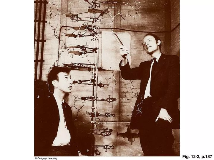
Fig. 12-2, p.187. Fig. 12-5, p.190. 2-nanometer diameter overall. 0.34-nanometer distance between each pair of bases. 3.4-nanometer length of each full twist of the double helix. In all respects shown here, the Watson–Crick model for DNA structure is consistent with the

E N D
2-nanometer diameter overall 0.34-nanometer distance between each pair of bases 3.4-nanometer length of each full twist of the double helix In all respects shown here, the Watson–Crick model for DNA structure is consistent with the known biochemical and x-ray diffraction data. The pattern of base pairing (A with T, and G with C) is consistent with the known composition of DNA (A = T, and G = C). Fig. 12-6, p.191
new old old new Fig. 12-8, p.192
Part of a parent DNA molecule, with two complementary strands of base-paired nucleotides. Replication starts. The strands are unwound at many sites along the molecule’s length. Each of the two parent strands guides the assembly of new DNA strands from free nucleotides, according to base-pairing rules. Any gaps between bases of the “new” DNA are joined to form a continuous strand. The base sequence of each half-old, half-new DNA molecule is identical to that of the parent. Fig. 12-9, p.193
newly forming RNA transcript gene region RNA polymerase, the enzyme that catalyzes transcription DNA template winding up DNA template unwinding Fig. 13-3, p.198
anticodon amino acid attachment site Fig. 13-7, p.201
Initiation A mature mRNA leaves the nucleus and enters cytoplasm, which has many free amino acids, tRNAs, and ribosome subunits. An initiator tRNA binds to a small ribosomal subunit and the mRNA. mRNA small ribosomal subunit initiator tRNA large ribosomal subunit A large ribosomal subunit joins, and the cluster is now called an initiation complex. Fig. 13-8, p.202
Elongation An initiator tRNA carries the amino acid methionine, so the first amino acid of the new polypeptide chain will be methionine. A second tRNA binds the second codon of the mRNA (here, that codon is GUG, so the tRNA that binds carries the amino acid valine). A peptide bond forms between the first two amino acids (here, methionine and valine). The first tRNA is released and the ribosome moves to the next codon in the mRNA. A third tRNA binds to the third codon of the mRNA (here, that codon is UUA, so the tRNA carries the amino acid leucine). A peptide bond forms between the second and third amino acids (here, valine and leucine). Fig. 13-8, p.202
The second RNA is released and the ribosome moves to the next codon. A fourth tRNA binds the fourth mRNA codon (here, that codon is GGG, so the tRNA carries the amino acid glycine). A peptide bond forms between the third and fourth amino acids (here, leucine and glycine) Termination Steps d and e are repeated over and over until the ribosome encounters a STOP codon in the mRNA. The mRNA transcript and the new polypeptide chain are released from the ribosome. The two ribosomal subunits separate from each other. Translation is now complete. Either the chain will join the pool of proteins in the cytoplasm or it will enter rough ER of the endomembrane system (Section 4.8). Fig. 13-8, p.202
part of DNA mRNA transcribed from DNA resulting amino acid sequence GLUTAMATE THREONINE PROLINE GLUTAMATE LYSINE base substitution in DNA altered mRNA altered amino acid sequence THREONINE PROLINE VALINE GLUTAMATE LYSINE deletion in DNA altered mRNA altered amino acid sequence THREONINE PROLINE GLYCINE ARGININE Fig. 13-9, p.202
Assembly of RNA on unwound regions of DNA molecule Transcription tRNA mRNA rRNA mRNA processing proteins ribosomal subunits mature tRNA mature mRNA transcripts Convergence of RNAs cytoplasmic pools of amino acids, ribosomal subunits, and tRNAs Translation At an intact ribosome, synthesis of a polypeptide chain at the binding sites for mRNA and tRNAs Final protein Fig. 13-11, p.206